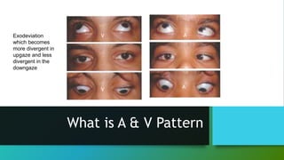
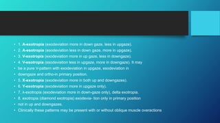
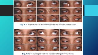
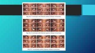
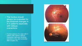
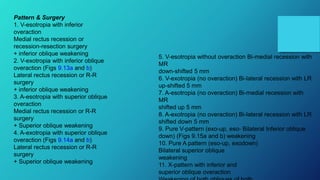
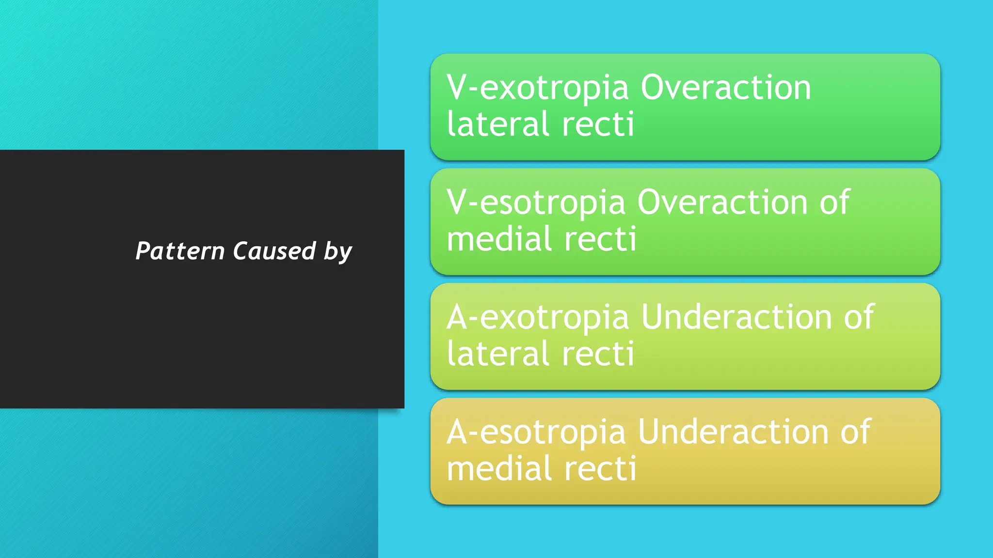
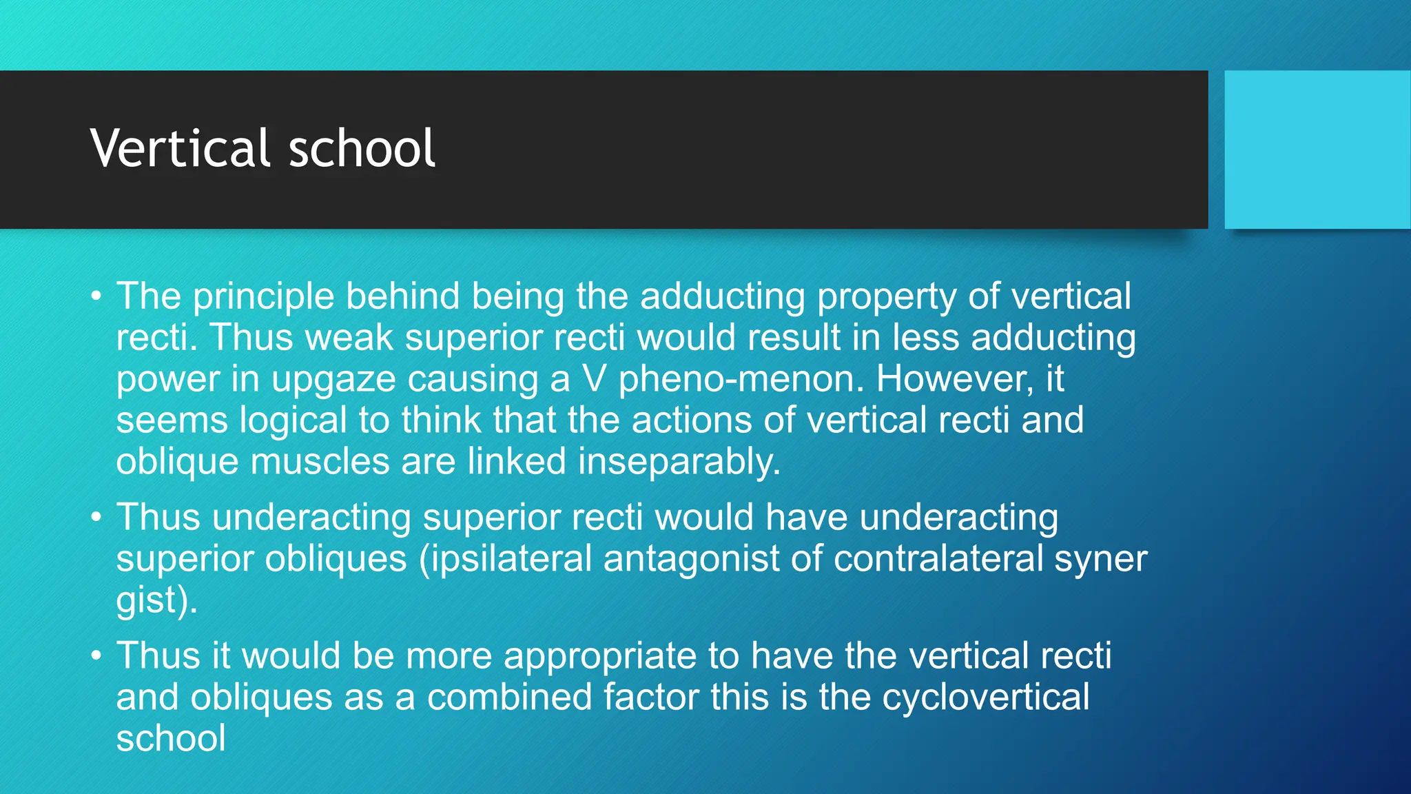
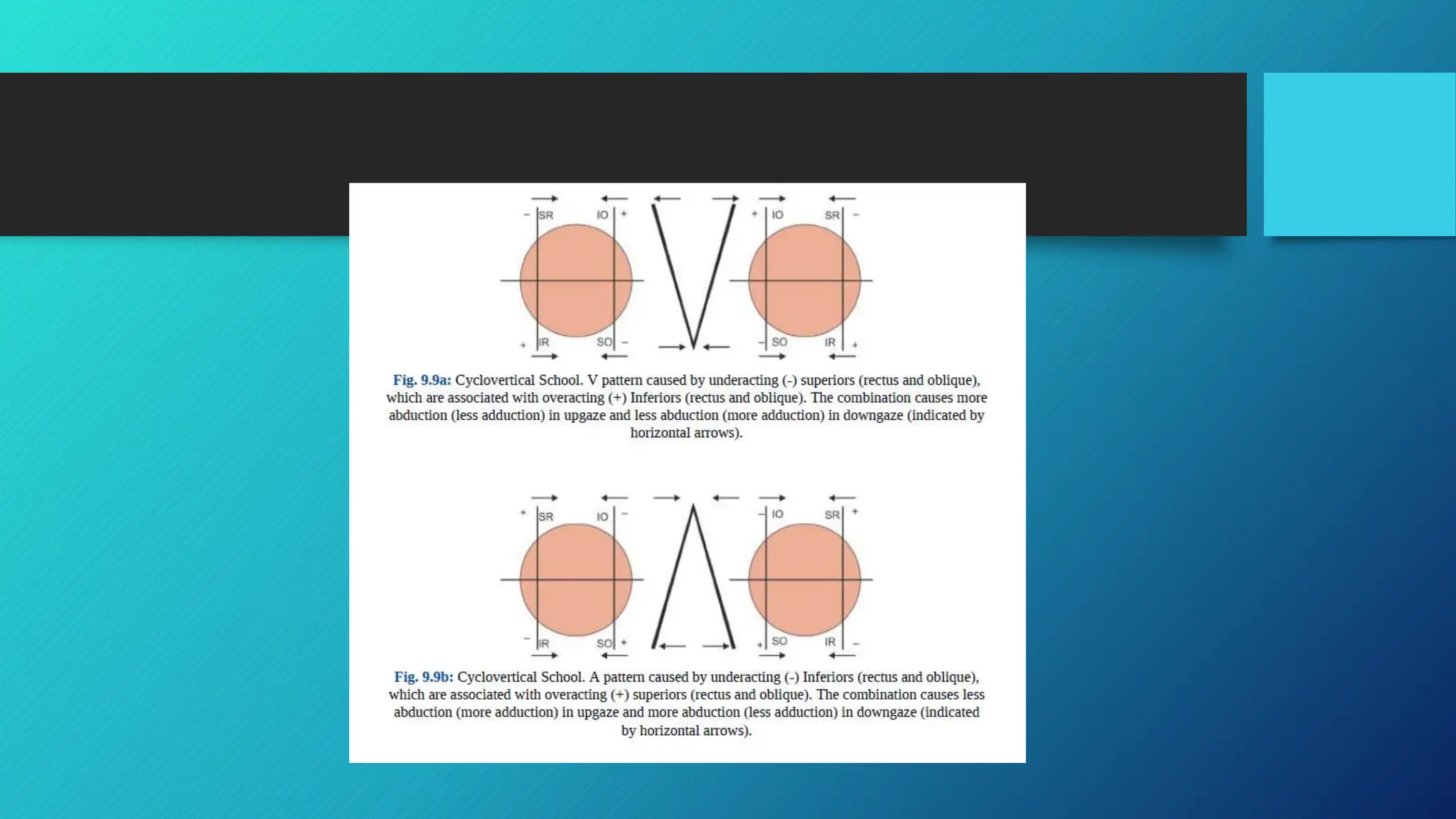
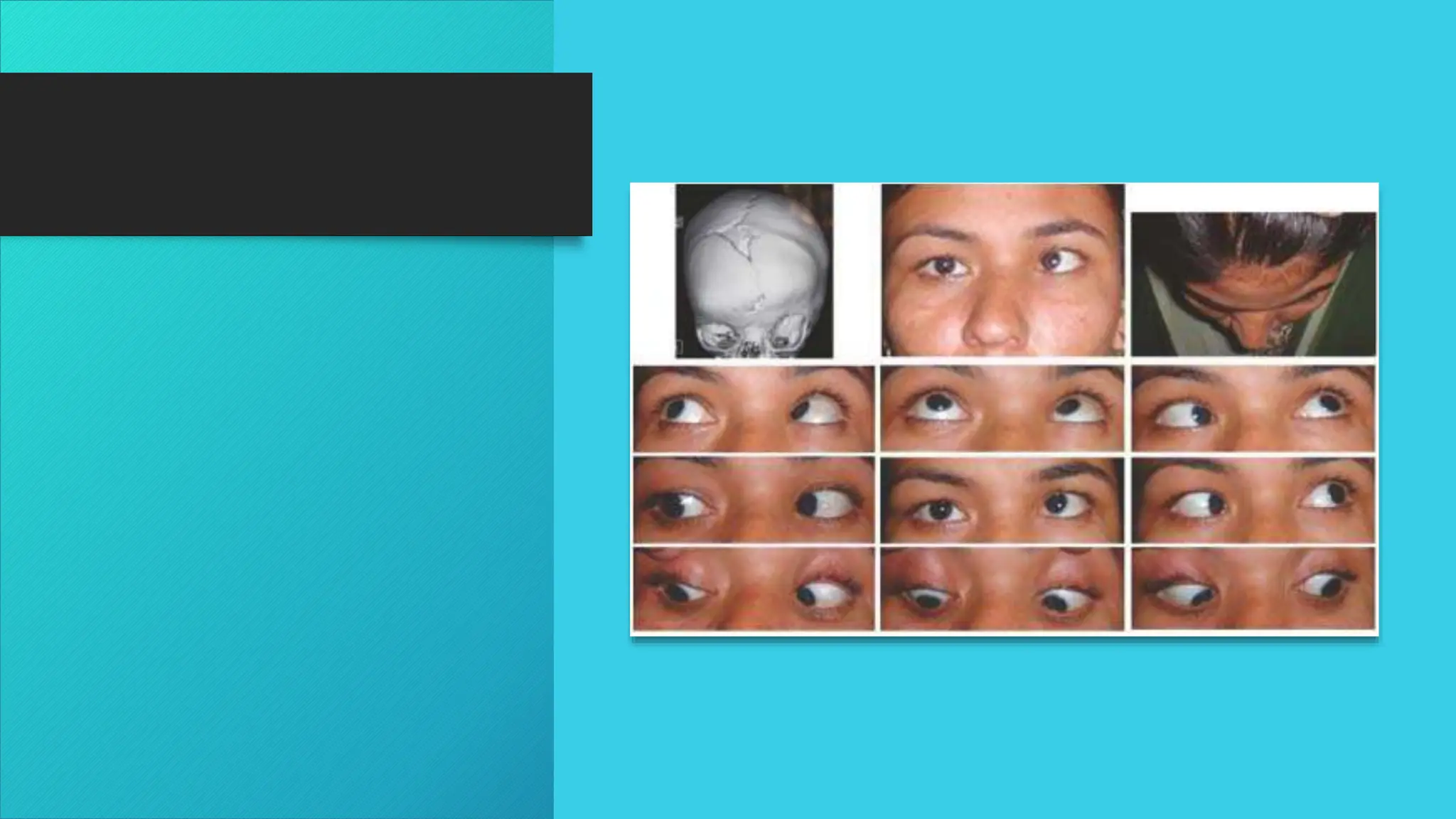
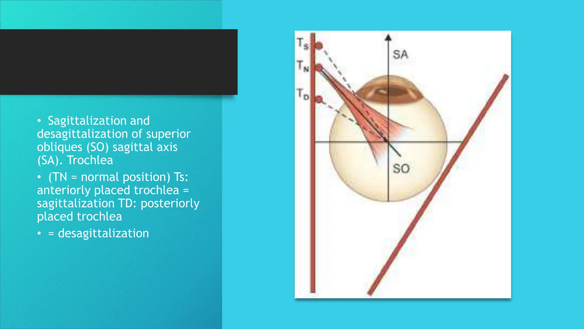
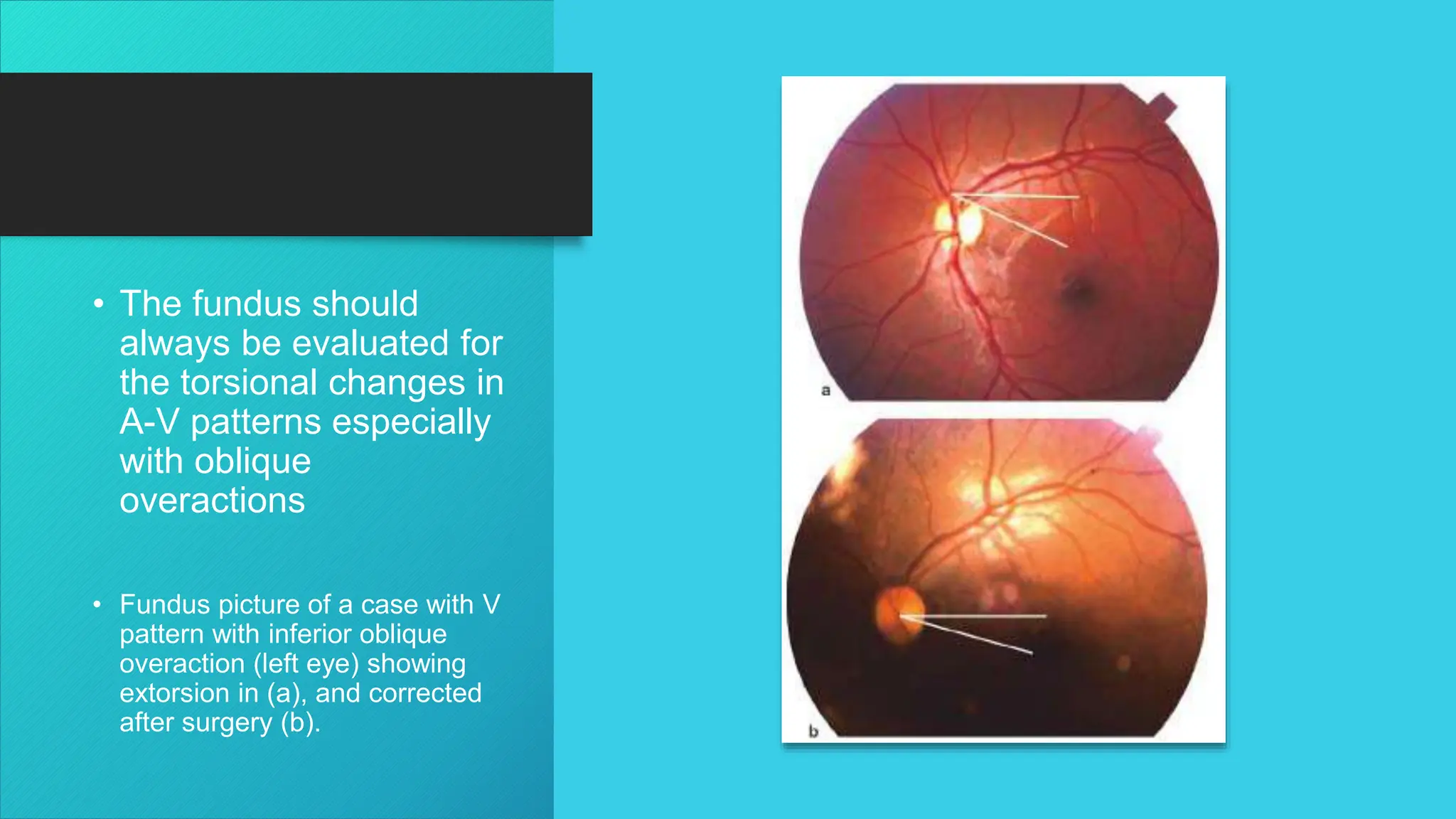
KEMBAR78
A – V PATTERNS : Introduction , Types & Management | PPTX
A – V PATTERNS
Jasmin
Context
Introduction
• Horizontal deviations are comitant in horizontal gaze but may not
in vertical gaze
• Vertical are not…..It is called as …..
vertical incomitant comitant horizontal deviation
• Introduced alphabets The latter is because the changes of
horizontal deviation in up- and downgaze resembles the alphabets
A or V.
What is A & V Pattern
Exodeviation
which becomes
more divergent in
upgaze and less
divergent in the
downgaze
V
V
A
A
• 1. A-exotropia (exodeviation more in down gaze, less in upgaze).
• 2. A-esotropia (esodeviation less in down gaze, more in upgaze).
• 3. V-exotropia (exodeviation more in up gaze, less in downgaze).
• 4. V-esotropia (esodeviation less in upgaze, more in downgaze). It may
• be a pure V-pattern with exodeviation in upgaze, esodeviation in
• downgaze and ortho-in primary position.
• 5. X-exotropia (exodeviation more in both up and downgazes).
• 6. Y-exotropia (exodeviation more in upgaze only).
• 7. λ-exotropia (exodeviation more in down-gaze only), delta exotropia.
• 8. exotropia (diamond exotropia) exodevia- tion only in primary position
• not in up and downgazes.
• Clinically these patterns may be present with or without oblique muscle overactions
Prevalance
• Since Urrets-Zavalia described them in 1948, A and V patterns have been commonly seen
in at least one-third of esotropia and exotropia.
• V patterns are more common than A patterns. While esodeviations are more common in
the West the exodeviations are more common in the Indian and the African races. The A-
esotropia is the least type of pattern amongst the four main types, otherwise the diamond
pattern is the least of all.
Etiology : Horizontal school
• Urist championed the role of horizontal recti, assuming that lateral
recti are more effective in upgaze and medial recti more effective in
downgaze. An overaction of lateral recti causes V-exotropia and
underaction of medial recti causes A-esotropia
• Cases with A-V patterns which do not have oblique muscle
overactions could be responsible because of this factor. Based on
this principle Urist advocated surgery to correct the specific
anomaly, i.e. in case of overaction of the recti, recess them and in
case of underaction resect them.
Pattern Caused by
V-exotropia Overaction
lateral recti
V-esotropia Overaction of
medial recti
A-exotropia Underaction of
lateral recti
A-esotropia Underaction of
medial recti
Vertical school
• The principle behind being the adducting property of vertical
recti. Thus weak superior recti would result in less adducting
power in upgaze causing a V pheno-menon. However, it
seems logical to think that the actions of vertical recti and
oblique muscles are linked inseparably.
• Thus underacting superior recti would have underacting
superior obliques (ipsilateral antagonist of contralateral syner
gist).
• Thus it would be more appropriate to have the vertical recti
and obliques as a combined factor this is the cyclovertical
school
Pattern
caused by
Inferior oblique Superior
oblique V pattern
Inferior rectus Superior rectus
V pattern
Superior oblique Inferior
oblique A pattern
SuperiorSuperior Inferior
rectus A pattern
Structural factors
• The trochlea acts as the functional origin of the superior oblique.
• The role of sagittalization: the oblique muscles becoming more parallel to the
sagittal (anteroposterior) axis
• Desagittalization: when the oblique muscles become more parallel to the
coronal plane have been stressed by Gobin. If the superior oblique is
desagittalised due to the retro placement of trochlea (as in plagiocephaly), it
becomes a poorer depressor. And relatively the inferior oblique becomes a
stronger elevator (Figs 9.10a and b).
• Similarly with a more frontally placed trochlea (as in hydrocephalus with frontal
bossing), superior oblique becomes more sagittalised in relation to the inferior
oblique making it a stronger elevator. This relative action of the obliques can
cause A and V patterns.
• Sagittalization and
desagittalization of superior
obliques (SO) sagittal axis
(SA). Trochlea
• (TN = normal position) Ts:
anteriorly placed trochlea =
sagittalization TD: posteriorly
placed trochlea
• = desagittalization
Diagnosis
• The horizontal deviations are measured in 25° upgaze and 35° downgaze in
addition to the primary position. Sometimes it may be desirable to check in 45°
downgaze position to exclude a significant A pattern.
• A 15 pd difference is taken as significant for a V pattern and a 10 pd difference
is taken as significant for A pattern. For repeatable and reproducible
measurements these positions are quantified with the help of a scale and a
protractor on the lateral side of the head. A 35° chin-up and 25° chin down
position is used with a 6 m distance target for distance fixation measurement. For
near fixation at 33 cm, either the head is similarly tilted or the target is suitably
fixed.
• A cephalodeviometer has been devised using a mirror with markings drawn on
it. The patient is made to wear a head band with a vertical marker, and head is
suitably tilted for the required position.
• Children with V-exotropia with BSV in downgaze may have a chin-up
posture and with A-esotropia with BSV in upgaze may have a chin down
• The fundus should
always be evaluated for
the torsional changes in
A-V patterns especially
with oblique
overactions
• Fundus picture of a case with V
pattern with inferior oblique
overaction (left eye) showing
extorsion in (a), and corrected
after surgery (b).
Treatment
5. V-esotropia without overaction Bi-medial recession with
MR
down-shifted 5 mm
6. V-exotropia (no overaction) Bi-lateral recession with LR
up-shifted 5 mm
7. A-esotropia (no overaction) Bi-medial recession with
MR
shifted up 5 mm
8. A-exotropia (no overaction) Bi-lateral recession with LR
shifted down 5 mm
9. Pure V-pattern (exo-up, eso- Bilateral Inferior oblique
down) (Figs 9.15a and b) weakening
10. Pure A pattern (eso-up, exodown)
Bilateral superior oblique
weakening
11. X-pattern with inferior and
superior oblique overaction
Pattern & Surgery
1. V-esotropia with inferior
overaction
Medial rectus recession or
recession-resection surgery
+ inferior oblique weakening
2. V-exotropia with inferior oblique
overaction (Figs 9.13a and b)
Lateral rectus recession or R-R
surgery
+ inferior oblique weakening
3. A-esotropia with superior oblique
overaction
Medial rectus recession or R-R
surgery
+ Superior oblique weakening
4. A-exotropia with superior oblique
overaction (Figs 9.14a and b)
Lateral rectus recession or R-R
surgery
+ Superior oblique weakening

A – V PATTERNS : Introduction , Types & Management

  • 1.
    A – VPATTERNS Jasmin
  • 2.
  • 3.
    Introduction • Horizontal deviationsare comitant in horizontal gaze but may not in vertical gaze • Vertical are not…..It is called as ….. vertical incomitant comitant horizontal deviation • Introduced alphabets The latter is because the changes of horizontal deviation in up- and downgaze resembles the alphabets A or V.
  • 4.
    What is A& V Pattern Exodeviation which becomes more divergent in upgaze and less divergent in the downgaze V V A A
  • 5.
    • 1. A-exotropia(exodeviation more in down gaze, less in upgaze). • 2. A-esotropia (esodeviation less in down gaze, more in upgaze). • 3. V-exotropia (exodeviation more in up gaze, less in downgaze). • 4. V-esotropia (esodeviation less in upgaze, more in downgaze). It may • be a pure V-pattern with exodeviation in upgaze, esodeviation in • downgaze and ortho-in primary position. • 5. X-exotropia (exodeviation more in both up and downgazes). • 6. Y-exotropia (exodeviation more in upgaze only). • 7. λ-exotropia (exodeviation more in down-gaze only), delta exotropia. • 8. exotropia (diamond exotropia) exodevia- tion only in primary position • not in up and downgazes. • Clinically these patterns may be present with or without oblique muscle overactions
  • 8.
    Prevalance • Since Urrets-Zavaliadescribed them in 1948, A and V patterns have been commonly seen in at least one-third of esotropia and exotropia. • V patterns are more common than A patterns. While esodeviations are more common in the West the exodeviations are more common in the Indian and the African races. The A- esotropia is the least type of pattern amongst the four main types, otherwise the diamond pattern is the least of all.
  • 9.
    Etiology : Horizontalschool • Urist championed the role of horizontal recti, assuming that lateral recti are more effective in upgaze and medial recti more effective in downgaze. An overaction of lateral recti causes V-exotropia and underaction of medial recti causes A-esotropia • Cases with A-V patterns which do not have oblique muscle overactions could be responsible because of this factor. Based on this principle Urist advocated surgery to correct the specific anomaly, i.e. in case of overaction of the recti, recess them and in case of underaction resect them.
  • 10.
    Pattern Caused by V-exotropiaOveraction lateral recti V-esotropia Overaction of medial recti A-exotropia Underaction of lateral recti A-esotropia Underaction of medial recti
  • 12.
    Vertical school • Theprinciple behind being the adducting property of vertical recti. Thus weak superior recti would result in less adducting power in upgaze causing a V pheno-menon. However, it seems logical to think that the actions of vertical recti and oblique muscles are linked inseparably. • Thus underacting superior recti would have underacting superior obliques (ipsilateral antagonist of contralateral syner gist). • Thus it would be more appropriate to have the vertical recti and obliques as a combined factor this is the cyclovertical school
  • 13.
    Pattern caused by Inferior obliqueSuperior oblique V pattern Inferior rectus Superior rectus V pattern Superior oblique Inferior oblique A pattern SuperiorSuperior Inferior rectus A pattern
  • 15.
    Structural factors • Thetrochlea acts as the functional origin of the superior oblique. • The role of sagittalization: the oblique muscles becoming more parallel to the sagittal (anteroposterior) axis • Desagittalization: when the oblique muscles become more parallel to the coronal plane have been stressed by Gobin. If the superior oblique is desagittalised due to the retro placement of trochlea (as in plagiocephaly), it becomes a poorer depressor. And relatively the inferior oblique becomes a stronger elevator (Figs 9.10a and b). • Similarly with a more frontally placed trochlea (as in hydrocephalus with frontal bossing), superior oblique becomes more sagittalised in relation to the inferior oblique making it a stronger elevator. This relative action of the obliques can cause A and V patterns.
  • 17.
    • Sagittalization and desagittalizationof superior obliques (SO) sagittal axis (SA). Trochlea • (TN = normal position) Ts: anteriorly placed trochlea = sagittalization TD: posteriorly placed trochlea • = desagittalization
  • 18.
    Diagnosis • The horizontaldeviations are measured in 25° upgaze and 35° downgaze in addition to the primary position. Sometimes it may be desirable to check in 45° downgaze position to exclude a significant A pattern. • A 15 pd difference is taken as significant for a V pattern and a 10 pd difference is taken as significant for A pattern. For repeatable and reproducible measurements these positions are quantified with the help of a scale and a protractor on the lateral side of the head. A 35° chin-up and 25° chin down position is used with a 6 m distance target for distance fixation measurement. For near fixation at 33 cm, either the head is similarly tilted or the target is suitably fixed. • A cephalodeviometer has been devised using a mirror with markings drawn on it. The patient is made to wear a head band with a vertical marker, and head is suitably tilted for the required position. • Children with V-exotropia with BSV in downgaze may have a chin-up posture and with A-esotropia with BSV in upgaze may have a chin down
  • 19.
    • The fundusshould always be evaluated for the torsional changes in A-V patterns especially with oblique overactions • Fundus picture of a case with V pattern with inferior oblique overaction (left eye) showing extorsion in (a), and corrected after surgery (b).
  • 20.
  • 21.
    5. V-esotropia withoutoveraction Bi-medial recession with MR down-shifted 5 mm 6. V-exotropia (no overaction) Bi-lateral recession with LR up-shifted 5 mm 7. A-esotropia (no overaction) Bi-medial recession with MR shifted up 5 mm 8. A-exotropia (no overaction) Bi-lateral recession with LR shifted down 5 mm 9. Pure V-pattern (exo-up, eso- Bilateral Inferior oblique down) (Figs 9.15a and b) weakening 10. Pure A pattern (eso-up, exodown) Bilateral superior oblique weakening 11. X-pattern with inferior and superior oblique overaction Pattern & Surgery 1. V-esotropia with inferior overaction Medial rectus recession or recession-resection surgery + inferior oblique weakening 2. V-exotropia with inferior oblique overaction (Figs 9.13a and b) Lateral rectus recession or R-R surgery + inferior oblique weakening 3. A-esotropia with superior oblique overaction Medial rectus recession or R-R surgery + Superior oblique weakening 4. A-exotropia with superior oblique overaction (Figs 9.14a and b) Lateral rectus recession or R-R surgery + Superior oblique weakening