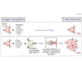
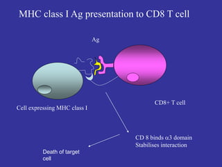
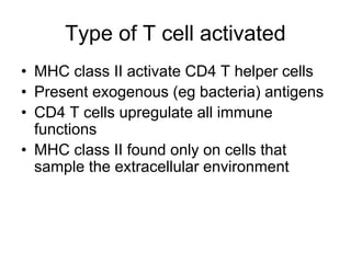
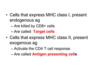
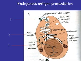
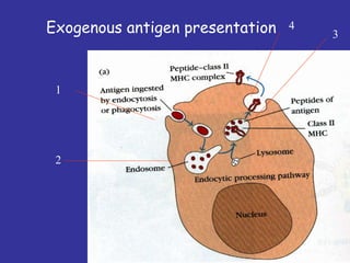
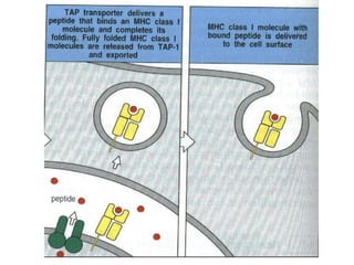
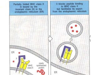
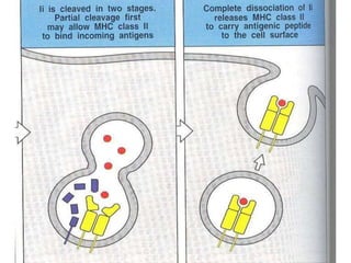
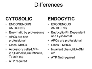
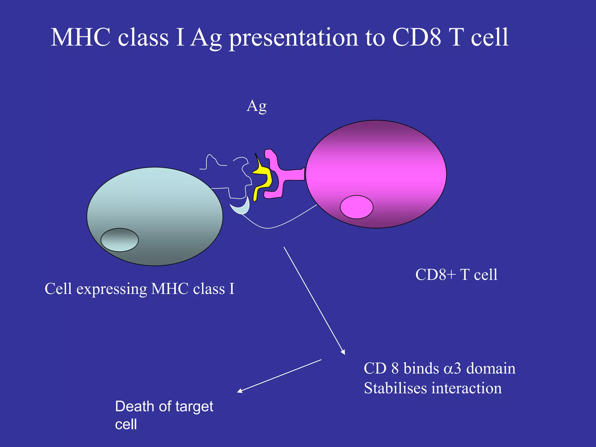
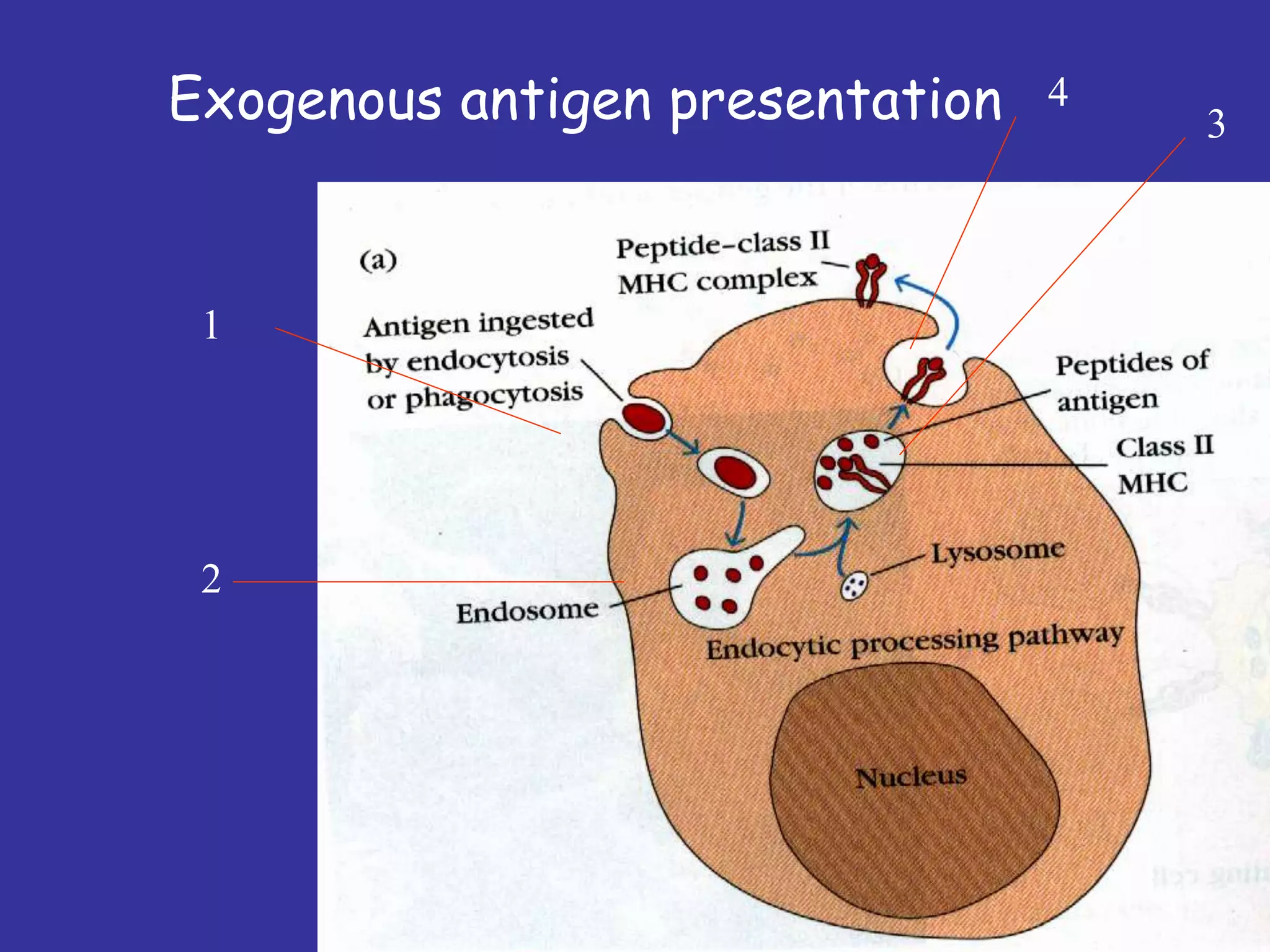
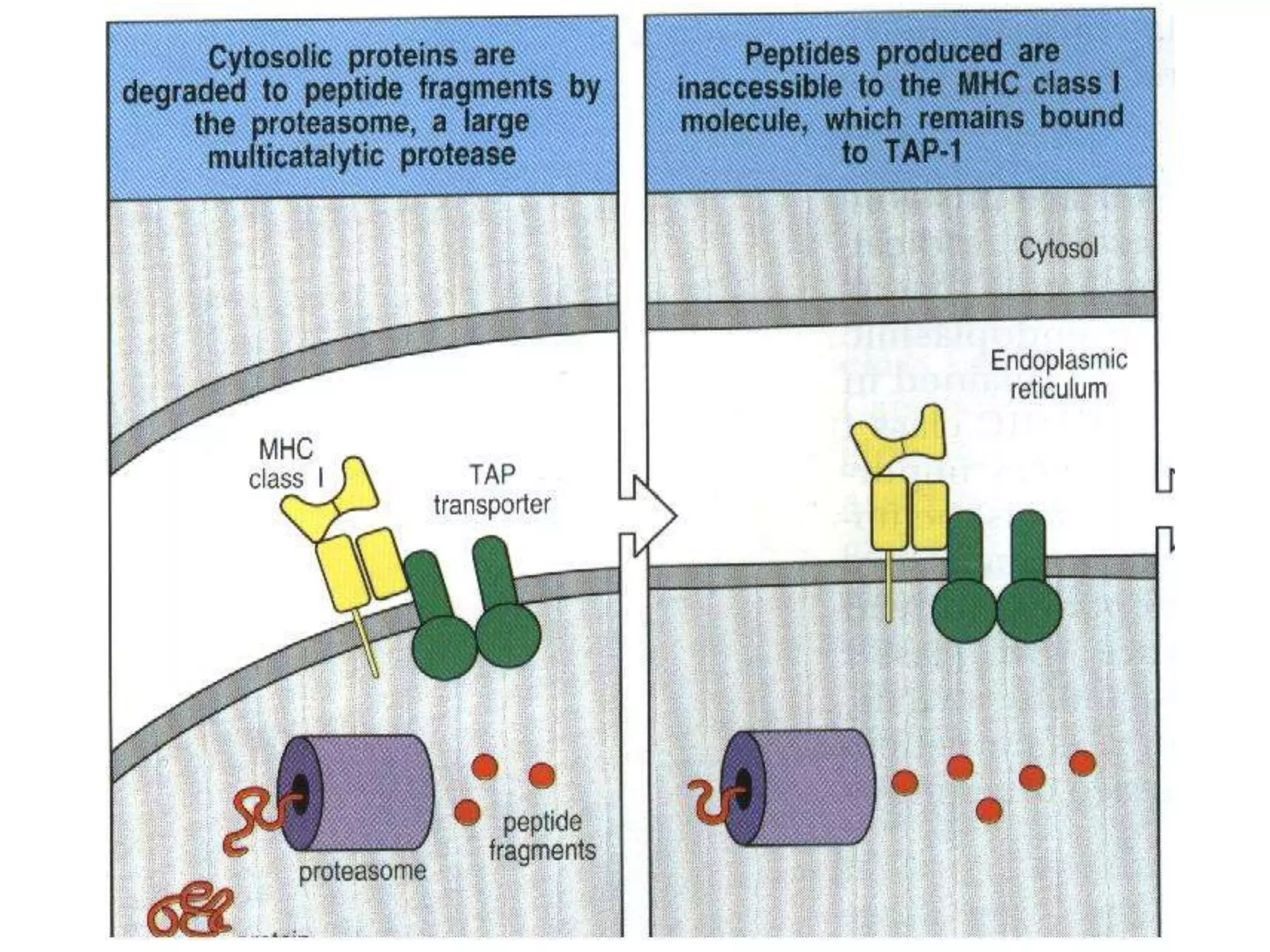
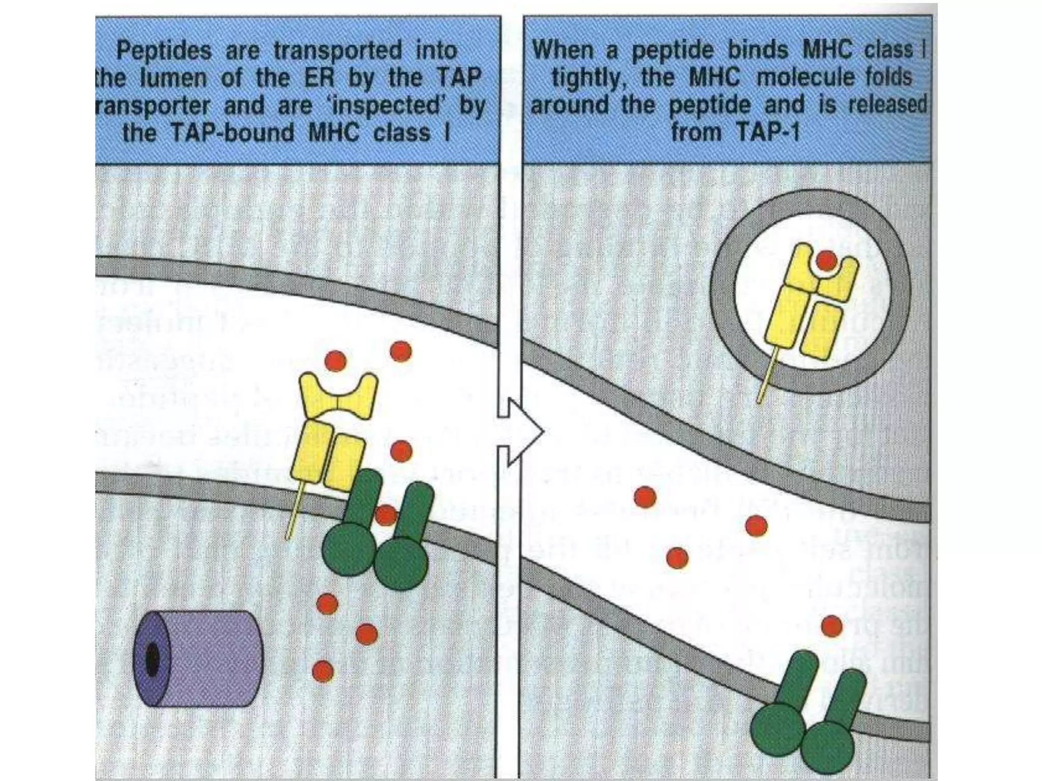
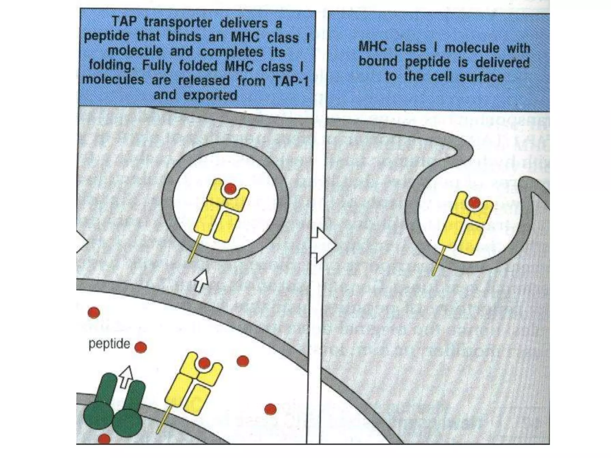
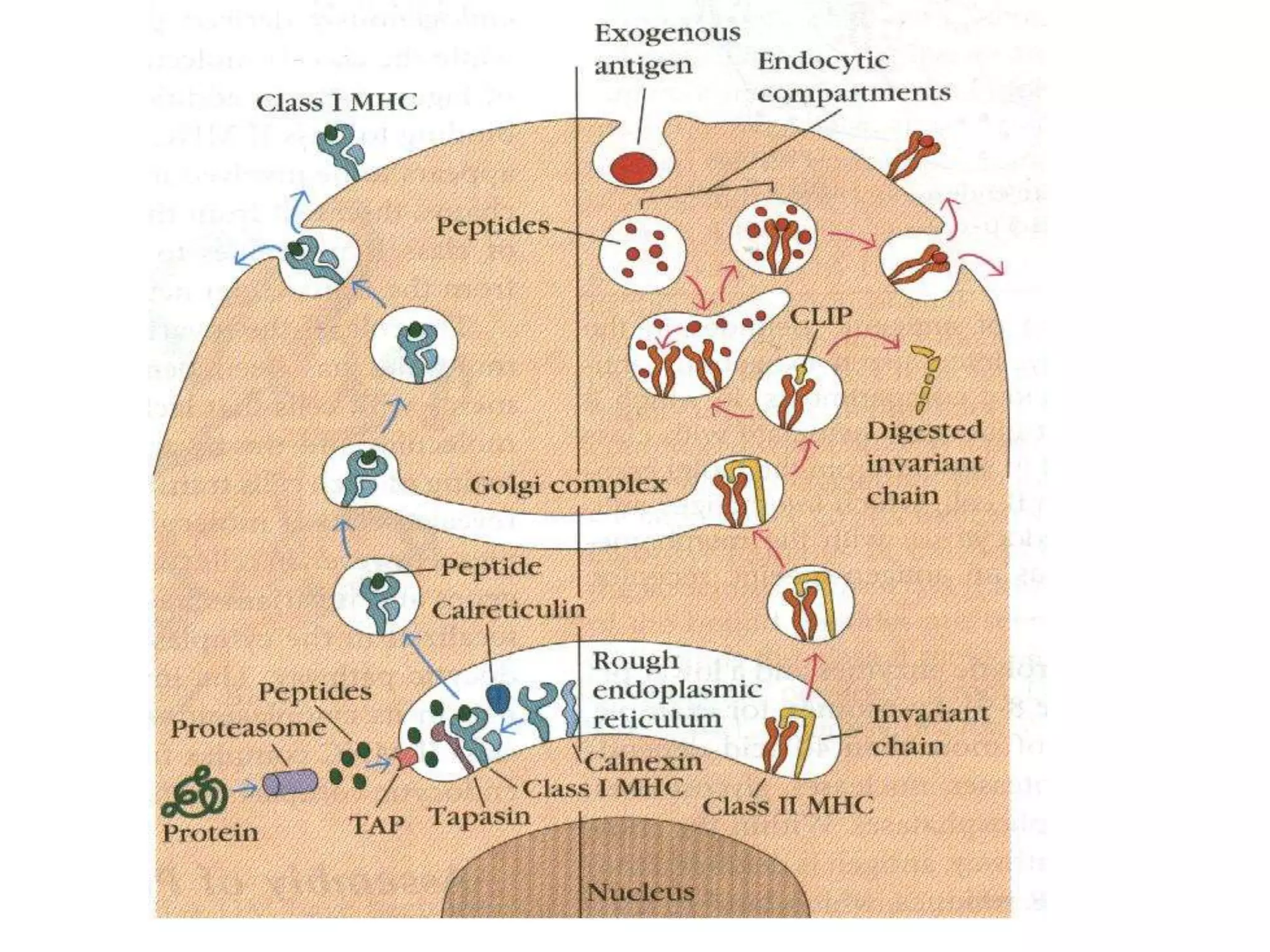
KEMBAR78
Antigen processing-and-presentation-09 | PPT
APC,Antigen processing and
presentation
Dr. Dinesh Jain
SMS MC Jaipur
Antigen processing and
presentation
• Why is it needed?
• How does it happen?
• How are the pathways of endogenous and
exogenous antigen kept apart?
• What are the consequences?
The Immune Response
Immune Response: Involves production
of antibodies and generation of
specialized lymphocytes against specific
antigens.
Antigen: Molecules from a pathogen or
foreign organism that provoke a specific
immune response.
The Immune System
Innate or Genetic Immunity: Immunity
an organism is born with.
– Genetically determined.
– May be due to lack of receptors or other
molecules required for infection.
• Immunity of mice to poliovirus.
Acquired Immunity: Immunity that an
organism develops during lifetime.
– Not genetically determined.
– May be acquired naturally or artificially.
• Development of immunity to measles in
response to infection or vaccination.
• Found in bone marrow
• Give rise to blood and immune cells
– Form two lineages: myeloid and lymphoid
Hematopoietic Stem Cells
Myeloid Lineage
• Gives rise to eight types of cells found in
body tissues, focusing on three
1. Mast Cells
• Degranulate hormonal mediators when activated,
involved with inflammation
• Activated by IgE antibodies, physical/chemical injury,
or complement proteins
• Morphology: typical round cellular shape
• Defining Organelles: secretory granules and lipid
bodies
Myeloid Lineage
2. Macrophages
• Matured monocytes that migrate into tissues from the
circulatory system
• Role is to phagocytize dead cells and pathogens
– Produce NO, O2
-, and H2O2 to break down material.
• Specific names for specific locations, ex: Kupffer cells
in liver ,alveolar in lungs ,mesangial in
kidneys.,microglial in brain
• Morphology: monocyte is a spherical shape, but
macrophage shape depends on what tissue it goes
and matures.
• Defining Organelles: extensive Rough ER,
lysosomes/phagosomes, pseudopods
3. Dendritic Cells
• Main role is to process antigen material and present it
to other cells on its surface.
• Antigen presenting cell
• Morphology: form branch like projections (dendrites)
during stage of development
• Defining Organelles: Golgi, ER,
(proteasome – protein complex that degrades
proteins)
Myeloid Lineage
• Gives rise to three major types of cells
1. Natural Killer Cells (NK)
• Role: limits the spread of tumors and microbial
infections by inducing apoptosis in cells, limiting
tissue damage
• Morphology: large granular lymphocyte,
spherical
• Defining Organelles: cytoplasmic lytic granules,
secretory lysosomes, golgi, vesicles,
microtubules
Lymphoid Lineage
2. B Cells
• Role: produce antibodies, become antigen-
presenting cells, become memory B cells
– Memory B cells “remember” specific antigens and
can launch fast immune response if antigen is
encountered again.
– Live for about 10 years.
• Morphology: spherical, with very large nucleus
and scant cytoplasm around it, has B-cell
receptor on cell surface
• Defining Organelles: large ER and Golgi,
lysosomes
Lymphoid Lineage
3. T Cells
• Several types including T Helper and cytotoxic
T cells
• Role: secrete cytokines after activation by APC
to help aid active immune responses, and
destroy virally infected and tumor cells by
binding to antigens on their surface,
respectively
• Morphology: Spherical with T-cell receptor on
cell surface, covered with short microvilli
• Defining Organelles: ER, Golgi
Lymphoid Lineage
Components of Human Immune
System
• I. Humoral (Antibody-Mediated) Immunity
– Involves production of antibodies
– Produced by a subset of lymphocytes called B
cells.
– B cells that are stimulated are called plasma
cells.
– found in extracellular fluids (blood plasma,
lymph, mucus, etc.) and the surface of B cells.
– against bacteria, bacterial toxins, and
viruses that circulate freely in body fluids,
before they enter cells.
– certain reactions against transplanted tissue.
Antibodies are Proteins that Recognize Specific
Antigens
II. Cell Mediated Immunity
– Involves specialized set of lymphocytes called T
cells that recognize foreign antigens on the
surface of cells, organisms, or tissues:
• Helper T cells
• Cytotoxic T cells
– T cells regulate proliferation and activity of other
cells of the immune system: B cells,
macrophages, neutrophils, etc.
– Defense against:
• Bacteria and viruses that are inside host cells and are
inaccessible to antibodies.
• Fungi, protozoa, and helminths
• Cancer cells
• Transplanted tissue
Cell Mediated Immunity is Carried Out by T Lymphocytes
Antigens
Most are proteins or large polysaccharides
from a foreign organism.
– Microbes: Capsules, cell walls, toxins, viral
capsids, flagella, etc.
– Nonmicrobes: Pollen, egg white , red blood cell
surface molecules, serum proteins, and surface
molecules from transplanted tissue.
Lipids and nucleic acids are only antigenic
when combined with proteins or
polysaccharides.
Molecular weight of 10,000 or higher.
– Hapten: Small foreign molecule that is not antigenic.
Must be coupled to a carrier molecule to be antigenic.
Once antibodies are formed they will recognize hapten.
Antigens
Epitope:
Small part of an antigen that interacts
with an antibody.
Any given antigen may have several
epitopes.
Each epitope is recognized by a
different antibody.
Epitopes: Antigen Regions that
Interact with Antibodies
Antibodies
Proteins that recognize and bind to a particular
antigen with very high specificity.
Made in response to exposure to the antigen.
One virus or microbe may have several antigenic
determinant sites, to which different antibodies
may bind.
Each antibody has at least two identical sites that
bind antigen: Antigen binding sites.
Valence of an antibody: Number of antigen
binding sites. Most are bivalent.
Belong to a group of serum proteins called
immunoglobulins (Igs).
Antibody Structure
Consequences of Antibody
Binding
Major Histocompatibility
Complex
• System of glycoproteins bound on cell
membrane which can be recognized by immune
system
• Genes coding MHC are localized on
chromosome 6, some of these genes are
extremely polymorphic (signs of Mendelian
heredity, codominancy, en bloc transfer)
Major histocompatibility complex
• Class I – HLA A,B,C (E,F,G)
a – expressed on the surface of all
nucleated human cells
– antigen presentation to Tc-
lymphocytes
• Class II – HLA DR, DP, DQ
– expressed on the surface of APC
(macrophages, B lymphocytes)
– antigen presentation to Th-
lymphocytes
Major histocompatibility complex
• Class III – HLA C2, C4, FB ( factor B), TNF-alfa
and beta)
– function – processing and transport of
T-lymphocyte epitopes
– heat-shock proteins
– inflammation mediators
Class 1MHC
• Comprised of 2 molecules
–  chain (45 kDa), transmembrane
– 2-microglobulin (12 kDa)
– Non-covalently associated with each other
• Association Of  Chain and 2 Is Required For
Surface Expression
•  Chain Made Up Of 3 Domains (1, 2 and 3)
• 2-microglobulin Similar To 3
• 1 And 2 Form Peptide Binding Cleft
– Fits peptide of about 8-10 a/a long
• 3 Highly Conserved Among MHC I Molecules
– Interacts with CD8 (TC) molecule
Class IIMHC
• Comprised of  and  chains
–  chain and  chain associate non-
covalently
•  and  chains made up of domains
– 1 and 2 ( chain)
– 1 and 2 ( chain)
• 1and 1 form antigen binding cleft
•  and  heterodimer has been shown to
dimerize
• CD4 molecule binds 2/2 domains
Antigen and epitopes
recognised by T and B cells
B cells
• Antigen/ epitope could be
proteins, lipids, nucleic
acids, polysaccharides
etc.
• surface conformational or
sequential
• Free state
• soluble
Tcells
• Proteins/ peptides mainly
• Surface linear or
sequential –deeply buried
• Only in combination with
MCH
• On cell surface
Antiagen presentation cells
required
1.
3.
2
TCR
MHC
Professional and non-professional
APCs
Professional APCs
• Dendritic cells
• Bcells
• Macrophages
Non-professional
APCs
• Fibroblasts(skin),Vascular
endothelial cells
• Glial cells (brain),Thymic
epithelial cells
• Pancreatic beta cells
• Dendritic cells are most efficient
because of high levels of MHC-II
molecules and B-7. B-7 binds with CD28
on T Helper cells.This has a
costimulatory signal.
• Macrophages need to be activated by
phagocytosis before MHCII and B-7
expression
Neutrophils
Neutrophils move through the blood to the site of infection.
Free to share, print, make copies and changes. Get yours at www.boundless.com
Wikipedia. "NeutrophilerAktion." CC BY-SA https://en.wikipedia.org/wiki/File:NeutrophilerAktion.png View on Boundless.com
Immunology
T cell by SEM
This scanning electron micrograph shows a T lymphocyte. T and B cells are indistinguishable by light microscopy, but can be differentiated
experimentally by probing their surface receptors.
Free to share, print, make copies and changes. Get yours at www.boundless.com
Connexions. "Adaptive Immunity." CC BY 3.0 http://cnx.org/content/m47434/latest/ View on Boundless.com
Immunology
Antigen presentation
• An antigen is a substance recognized by
immune system that reacts to its presence.
• For induction of specific immune response to
antigen, first of all antigen processing and its
presentation to APC is necessary.
• The professional antigen presenting cells
(APC) are cells expriming MHC class II
molecules (macrophages, dendritic cells, B-
lymphocytes).
Processing and presentation
of protein antigens
• 1/ Exogenous antigens
• Bacterial, helminthic or viral antigens
(either if they form immune complexes
swallowed by APC, or if they are
processed together with infected cells)
• They are presented in a complex with
MHC class II to T helper (CD4+) cells
Processing and presentation
of protein antigens
• 2/ Endogenous antigens
• Intracellular auto-antigens, antigens of
viruses or other intracellular parasites
(infecting APC) or tumorous antigens
• Present in complex with MHC class I
molecules to cytotoxic (CD8+) T cells
Type of T cell activated
• MHC class I activate CD8 T cytotoxic cells
• Present endogenous (eg viral) antigens ell
• Any cell can become virally
infected/neoplastic
• Therefore: MHC class I is on all nucleated
cells
Cell expressing MHC class I
Ag
CD8+ T cell
CD 8 binds 3 domain
Stabilises interaction
MHC class I Ag presentation to CD8 T cell
Death of target
cell
Type of T cell activated
• MHC class II activate CD4 T helper cells
• Present exogenous (eg bacteria) antigens
• CD4 T cells upregulate all immune
functions
• MHC class II found only on cells that
sample the extracellular environment
MHC class II-CD4 T cell
activation
• CD4 T helper cells up regulate immune
function
• Stimulate M activation and phagocytosis,
B cell antibody production
• Help to clear exogenous antigen
CD4+ T cell
CD 4 binds mainly 2 domain
Stabilises interaction
Cell expressing MHC class II
MHC class II Ag presentation to CD4 T cell
Upreglation of
Immune responses
B cell receptors
B cell receptors are embedded in the membranes of B cells and bind a variety of antigens through their variable regions, or antibodies. The signal
transduction region transfers the signal into the cell.
Free to share, print, make copies and changes. Get yours at www.boundless.com
Connexions. "Adaptive Immune Response." CC BY 3.0 http://cnx.org/content/m44821/latest/ View on Boundless.com
Immunology
Antigen: linear
Protein fragments
How does antigen get
Into MHC binding cleft
How does endogenous
Ag bind MHC class I?
How does exogenous ag
Bind MHC class II?
• Cells that express MHC class I, present
endogenous ag
– Are killed by CD8+ cells
– Are called Target cells
• Cells that express MHC class II, present
exogenous ag
– Activate the CD4 T cell response
– Are called Antigen presenting cells
Antigen processing and
presentation
• Separate pathways for endogenous and
exogenous antigens
• CYTOSOLIC pathway for endogenous
antigens
• ENDOCYTIC pathway for exogenous
antigens
Endogenous antigen presentation
1
2
3
Exogenous antigen presentation
1
2
3
4
Exogenous antigen presentation
1
2
3
4
Loading of antigen to MHC
class I
Loading of antigen to MHC
class II
Differences
CYTOSOLIC
• ENDOGENOUS
ANTIGENS
• Enzymatic by proteosome
• APCs are non
professional
• ClassI MHCs
• Accessory cells-LMP-
2,7,Calnexin,Calreticulin,
Tapsin etc
• ATP required
ENDOCYTIC
• EXOGENOUS
ANTIGENS
• Endocytic-Ph Dependent
and Lysosomal
• APCs are professional
• Class II MHCs
• Invariant chain,HLA-DM
etc
• ATP Not required
T Cells and Cell Mediated Immunity
Types of T cells
1. T Helper (TH) Cells: Central role in immune
response.
• Most are CD4+
• Recognize antigen on the surface of antigen
presenting cells (e.g.: macrophage).
• Activate macrophages
• Induce formation of cytotoxic T cells
• Stimulate B cells to produce antibodies.
Central Role of Helper T Cells
2. Cytotoxic T (Tc) Cells: Destroy
target cells.
• Most are CD4 negative (CD4 -).
• Recognize antigens on the surface of all
cells:
– Kill host cells that are infected with viruses or
bacteria.
– Recognize and kill cancer cells.
– Recognize and destroy transplanted tissue.
• Release protein called perforin which forms a
pore in target cell, causing lysis of infected
cells.
• Undergo apoptosis when stimulating antigen
is gone.
Cytotoxic T Cells Lyse Infected
Cells
3. Delayed Hypersensitivity T (TD) Cells:
Mostly T helper and a few cytotoxic T cells
that are involved in some allergic reactions
(poison ivy) and rejection of transplanted
tissue.
4. T Suppressor (Ts) Cells: May shut down
immune response.
Overview of the Immune
Response
Processing of non peptide
antigen
•CD1, A NON CLASSICAL MHC1
MOLECULE is involved
•Encoded by 5 GENES
(CD1A,CD1B,CD1C,CD1D,CD1E)on
chromosome 1
•Active on TAP-deficient cells also
BLS Bare lymphocyte
syndrome
•Low or absent expression of MHC
molecules on cells
•Type 1 BLS –defectiveTAP-1 and
TAP-2 Gene expression
• Gene therapy treatment
Useful textbooks
• Kuby Immunology
• Cellular and molecular immunology
(Abbas and Lichtman)
• Janeway’s Immunobiology (Murphy,
Travers, Walport)

Antigen processing-and-presentation-09

  • 1.
  • 2.
    Antigen processing and presentation •Why is it needed? • How does it happen? • How are the pathways of endogenous and exogenous antigen kept apart? • What are the consequences?
  • 3.
    The Immune Response ImmuneResponse: Involves production of antibodies and generation of specialized lymphocytes against specific antigens. Antigen: Molecules from a pathogen or foreign organism that provoke a specific immune response.
  • 4.
  • 5.
    Innate or GeneticImmunity: Immunity an organism is born with. – Genetically determined. – May be due to lack of receptors or other molecules required for infection. • Immunity of mice to poliovirus. Acquired Immunity: Immunity that an organism develops during lifetime. – Not genetically determined. – May be acquired naturally or artificially. • Development of immunity to measles in response to infection or vaccination.
  • 6.
    • Found inbone marrow • Give rise to blood and immune cells – Form two lineages: myeloid and lymphoid Hematopoietic Stem Cells
  • 7.
    Myeloid Lineage • Givesrise to eight types of cells found in body tissues, focusing on three 1. Mast Cells • Degranulate hormonal mediators when activated, involved with inflammation • Activated by IgE antibodies, physical/chemical injury, or complement proteins • Morphology: typical round cellular shape • Defining Organelles: secretory granules and lipid bodies
  • 8.
    Myeloid Lineage 2. Macrophages •Matured monocytes that migrate into tissues from the circulatory system • Role is to phagocytize dead cells and pathogens – Produce NO, O2 -, and H2O2 to break down material. • Specific names for specific locations, ex: Kupffer cells in liver ,alveolar in lungs ,mesangial in kidneys.,microglial in brain • Morphology: monocyte is a spherical shape, but macrophage shape depends on what tissue it goes and matures. • Defining Organelles: extensive Rough ER, lysosomes/phagosomes, pseudopods
  • 9.
    3. Dendritic Cells •Main role is to process antigen material and present it to other cells on its surface. • Antigen presenting cell • Morphology: form branch like projections (dendrites) during stage of development • Defining Organelles: Golgi, ER, (proteasome – protein complex that degrades proteins) Myeloid Lineage
  • 10.
    • Gives riseto three major types of cells 1. Natural Killer Cells (NK) • Role: limits the spread of tumors and microbial infections by inducing apoptosis in cells, limiting tissue damage • Morphology: large granular lymphocyte, spherical • Defining Organelles: cytoplasmic lytic granules, secretory lysosomes, golgi, vesicles, microtubules Lymphoid Lineage
  • 11.
    2. B Cells •Role: produce antibodies, become antigen- presenting cells, become memory B cells – Memory B cells “remember” specific antigens and can launch fast immune response if antigen is encountered again. – Live for about 10 years. • Morphology: spherical, with very large nucleus and scant cytoplasm around it, has B-cell receptor on cell surface • Defining Organelles: large ER and Golgi, lysosomes Lymphoid Lineage
  • 12.
    3. T Cells •Several types including T Helper and cytotoxic T cells • Role: secrete cytokines after activation by APC to help aid active immune responses, and destroy virally infected and tumor cells by binding to antigens on their surface, respectively • Morphology: Spherical with T-cell receptor on cell surface, covered with short microvilli • Defining Organelles: ER, Golgi Lymphoid Lineage
  • 13.
    Components of HumanImmune System
  • 14.
    • I. Humoral(Antibody-Mediated) Immunity – Involves production of antibodies – Produced by a subset of lymphocytes called B cells. – B cells that are stimulated are called plasma cells. – found in extracellular fluids (blood plasma, lymph, mucus, etc.) and the surface of B cells. – against bacteria, bacterial toxins, and viruses that circulate freely in body fluids, before they enter cells. – certain reactions against transplanted tissue.
  • 15.
    Antibodies are Proteinsthat Recognize Specific Antigens
  • 16.
    II. Cell MediatedImmunity – Involves specialized set of lymphocytes called T cells that recognize foreign antigens on the surface of cells, organisms, or tissues: • Helper T cells • Cytotoxic T cells – T cells regulate proliferation and activity of other cells of the immune system: B cells, macrophages, neutrophils, etc. – Defense against: • Bacteria and viruses that are inside host cells and are inaccessible to antibodies. • Fungi, protozoa, and helminths • Cancer cells • Transplanted tissue
  • 17.
    Cell Mediated Immunityis Carried Out by T Lymphocytes
  • 18.
    Antigens Most are proteinsor large polysaccharides from a foreign organism. – Microbes: Capsules, cell walls, toxins, viral capsids, flagella, etc. – Nonmicrobes: Pollen, egg white , red blood cell surface molecules, serum proteins, and surface molecules from transplanted tissue. Lipids and nucleic acids are only antigenic when combined with proteins or polysaccharides. Molecular weight of 10,000 or higher. – Hapten: Small foreign molecule that is not antigenic. Must be coupled to a carrier molecule to be antigenic. Once antibodies are formed they will recognize hapten.
  • 19.
    Antigens Epitope: Small part ofan antigen that interacts with an antibody. Any given antigen may have several epitopes. Each epitope is recognized by a different antibody.
  • 20.
    Epitopes: Antigen Regionsthat Interact with Antibodies
  • 21.
    Antibodies Proteins that recognizeand bind to a particular antigen with very high specificity. Made in response to exposure to the antigen. One virus or microbe may have several antigenic determinant sites, to which different antibodies may bind. Each antibody has at least two identical sites that bind antigen: Antigen binding sites. Valence of an antibody: Number of antigen binding sites. Most are bivalent. Belong to a group of serum proteins called immunoglobulins (Igs).
  • 22.
  • 23.
  • 24.
    Major Histocompatibility Complex • Systemof glycoproteins bound on cell membrane which can be recognized by immune system • Genes coding MHC are localized on chromosome 6, some of these genes are extremely polymorphic (signs of Mendelian heredity, codominancy, en bloc transfer)
  • 25.
    Major histocompatibility complex •Class I – HLA A,B,C (E,F,G) a – expressed on the surface of all nucleated human cells – antigen presentation to Tc- lymphocytes • Class II – HLA DR, DP, DQ – expressed on the surface of APC (macrophages, B lymphocytes) – antigen presentation to Th- lymphocytes
  • 26.
    Major histocompatibility complex •Class III – HLA C2, C4, FB ( factor B), TNF-alfa and beta) – function – processing and transport of T-lymphocyte epitopes – heat-shock proteins – inflammation mediators
  • 27.
    Class 1MHC • Comprisedof 2 molecules –  chain (45 kDa), transmembrane – 2-microglobulin (12 kDa) – Non-covalently associated with each other • Association Of  Chain and 2 Is Required For Surface Expression •  Chain Made Up Of 3 Domains (1, 2 and 3) • 2-microglobulin Similar To 3 • 1 And 2 Form Peptide Binding Cleft – Fits peptide of about 8-10 a/a long • 3 Highly Conserved Among MHC I Molecules – Interacts with CD8 (TC) molecule
  • 29.
    Class IIMHC • Comprisedof  and  chains –  chain and  chain associate non- covalently •  and  chains made up of domains – 1 and 2 ( chain) – 1 and 2 ( chain) • 1and 1 form antigen binding cleft •  and  heterodimer has been shown to dimerize • CD4 molecule binds 2/2 domains
  • 31.
    Antigen and epitopes recognisedby T and B cells B cells • Antigen/ epitope could be proteins, lipids, nucleic acids, polysaccharides etc. • surface conformational or sequential • Free state • soluble Tcells • Proteins/ peptides mainly • Surface linear or sequential –deeply buried • Only in combination with MCH • On cell surface
  • 32.
  • 33.
  • 34.
  • 35.
    Professional and non-professional APCs ProfessionalAPCs • Dendritic cells • Bcells • Macrophages Non-professional APCs • Fibroblasts(skin),Vascular endothelial cells • Glial cells (brain),Thymic epithelial cells • Pancreatic beta cells
  • 36.
    • Dendritic cellsare most efficient because of high levels of MHC-II molecules and B-7. B-7 binds with CD28 on T Helper cells.This has a costimulatory signal. • Macrophages need to be activated by phagocytosis before MHCII and B-7 expression
  • 37.
    Neutrophils Neutrophils move throughthe blood to the site of infection. Free to share, print, make copies and changes. Get yours at www.boundless.com Wikipedia. "NeutrophilerAktion." CC BY-SA https://en.wikipedia.org/wiki/File:NeutrophilerAktion.png View on Boundless.com Immunology
  • 38.
    T cell bySEM This scanning electron micrograph shows a T lymphocyte. T and B cells are indistinguishable by light microscopy, but can be differentiated experimentally by probing their surface receptors. Free to share, print, make copies and changes. Get yours at www.boundless.com Connexions. "Adaptive Immunity." CC BY 3.0 http://cnx.org/content/m47434/latest/ View on Boundless.com Immunology
  • 39.
    Antigen presentation • Anantigen is a substance recognized by immune system that reacts to its presence. • For induction of specific immune response to antigen, first of all antigen processing and its presentation to APC is necessary. • The professional antigen presenting cells (APC) are cells expriming MHC class II molecules (macrophages, dendritic cells, B- lymphocytes).
  • 40.
    Processing and presentation ofprotein antigens • 1/ Exogenous antigens • Bacterial, helminthic or viral antigens (either if they form immune complexes swallowed by APC, or if they are processed together with infected cells) • They are presented in a complex with MHC class II to T helper (CD4+) cells
  • 41.
    Processing and presentation ofprotein antigens • 2/ Endogenous antigens • Intracellular auto-antigens, antigens of viruses or other intracellular parasites (infecting APC) or tumorous antigens • Present in complex with MHC class I molecules to cytotoxic (CD8+) T cells
  • 42.
    Type of Tcell activated • MHC class I activate CD8 T cytotoxic cells • Present endogenous (eg viral) antigens ell • Any cell can become virally infected/neoplastic • Therefore: MHC class I is on all nucleated cells
  • 43.
    Cell expressing MHCclass I Ag CD8+ T cell CD 8 binds 3 domain Stabilises interaction MHC class I Ag presentation to CD8 T cell Death of target cell
  • 44.
    Type of Tcell activated • MHC class II activate CD4 T helper cells • Present exogenous (eg bacteria) antigens • CD4 T cells upregulate all immune functions • MHC class II found only on cells that sample the extracellular environment
  • 45.
    MHC class II-CD4T cell activation • CD4 T helper cells up regulate immune function • Stimulate M activation and phagocytosis, B cell antibody production • Help to clear exogenous antigen
  • 46.
    CD4+ T cell CD4 binds mainly 2 domain Stabilises interaction Cell expressing MHC class II MHC class II Ag presentation to CD4 T cell Upreglation of Immune responses
  • 47.
    B cell receptors Bcell receptors are embedded in the membranes of B cells and bind a variety of antigens through their variable regions, or antibodies. The signal transduction region transfers the signal into the cell. Free to share, print, make copies and changes. Get yours at www.boundless.com Connexions. "Adaptive Immune Response." CC BY 3.0 http://cnx.org/content/m44821/latest/ View on Boundless.com Immunology
  • 48.
    Antigen: linear Protein fragments Howdoes antigen get Into MHC binding cleft How does endogenous Ag bind MHC class I? How does exogenous ag Bind MHC class II?
  • 49.
    • Cells thatexpress MHC class I, present endogenous ag – Are killed by CD8+ cells – Are called Target cells • Cells that express MHC class II, present exogenous ag – Activate the CD4 T cell response – Are called Antigen presenting cells
  • 50.
    Antigen processing and presentation •Separate pathways for endogenous and exogenous antigens • CYTOSOLIC pathway for endogenous antigens • ENDOCYTIC pathway for exogenous antigens
  • 51.
  • 52.
  • 53.
  • 54.
    Loading of antigento MHC class I
  • 59.
    Loading of antigento MHC class II
  • 63.
    Differences CYTOSOLIC • ENDOGENOUS ANTIGENS • Enzymaticby proteosome • APCs are non professional • ClassI MHCs • Accessory cells-LMP- 2,7,Calnexin,Calreticulin, Tapsin etc • ATP required ENDOCYTIC • EXOGENOUS ANTIGENS • Endocytic-Ph Dependent and Lysosomal • APCs are professional • Class II MHCs • Invariant chain,HLA-DM etc • ATP Not required
  • 64.
    T Cells andCell Mediated Immunity Types of T cells 1. T Helper (TH) Cells: Central role in immune response. • Most are CD4+ • Recognize antigen on the surface of antigen presenting cells (e.g.: macrophage). • Activate macrophages • Induce formation of cytotoxic T cells • Stimulate B cells to produce antibodies.
  • 65.
    Central Role ofHelper T Cells
  • 66.
    2. Cytotoxic T(Tc) Cells: Destroy target cells. • Most are CD4 negative (CD4 -). • Recognize antigens on the surface of all cells: – Kill host cells that are infected with viruses or bacteria. – Recognize and kill cancer cells. – Recognize and destroy transplanted tissue. • Release protein called perforin which forms a pore in target cell, causing lysis of infected cells. • Undergo apoptosis when stimulating antigen is gone.
  • 67.
    Cytotoxic T CellsLyse Infected Cells
  • 68.
    3. Delayed HypersensitivityT (TD) Cells: Mostly T helper and a few cytotoxic T cells that are involved in some allergic reactions (poison ivy) and rejection of transplanted tissue. 4. T Suppressor (Ts) Cells: May shut down immune response.
  • 69.
    Overview of theImmune Response
  • 70.
    Processing of nonpeptide antigen •CD1, A NON CLASSICAL MHC1 MOLECULE is involved •Encoded by 5 GENES (CD1A,CD1B,CD1C,CD1D,CD1E)on chromosome 1 •Active on TAP-deficient cells also
  • 71.
    BLS Bare lymphocyte syndrome •Lowor absent expression of MHC molecules on cells •Type 1 BLS –defectiveTAP-1 and TAP-2 Gene expression • Gene therapy treatment
  • 72.
    Useful textbooks • KubyImmunology • Cellular and molecular immunology (Abbas and Lichtman) • Janeway’s Immunobiology (Murphy, Travers, Walport)