Image segmentation refers to partitioning a digital image into multiple regions or sets of pixels based on characteristics like color or texture. The goal is to simplify the image representation to make it easier to analyze. Some applications in medical imaging include locating tumors, measuring tissue volumes, and computer-guided surgery. Common segmentation techniques include thresholding, edge detection, region growing, and split-and-merge approaches.
Image segmentation divides images into meaningful regions for analysis, locating objects. Techniques include thresholding for foreground-background separation.
Edge detection highlights intensity contrasts, preserving key image structures. Thresholding distinguishes foreground from background based on intensity.
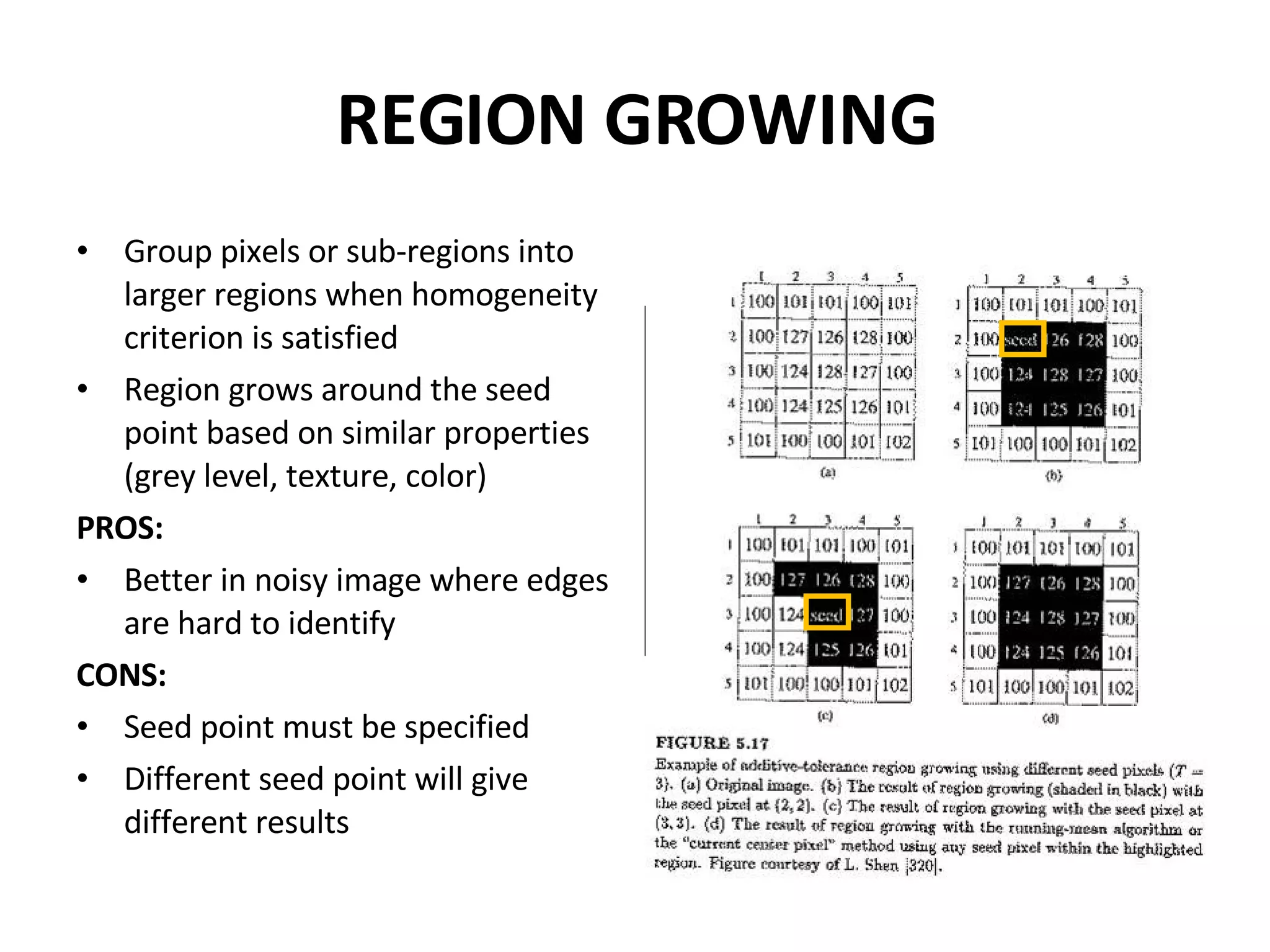
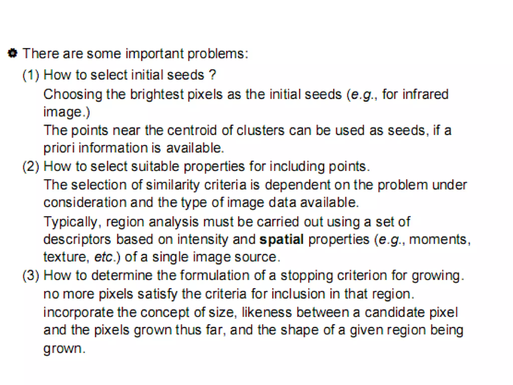
Region growing groups similar pixels for segmentation, aiding in noisy images. It requires seed points for homogeneity assessment.
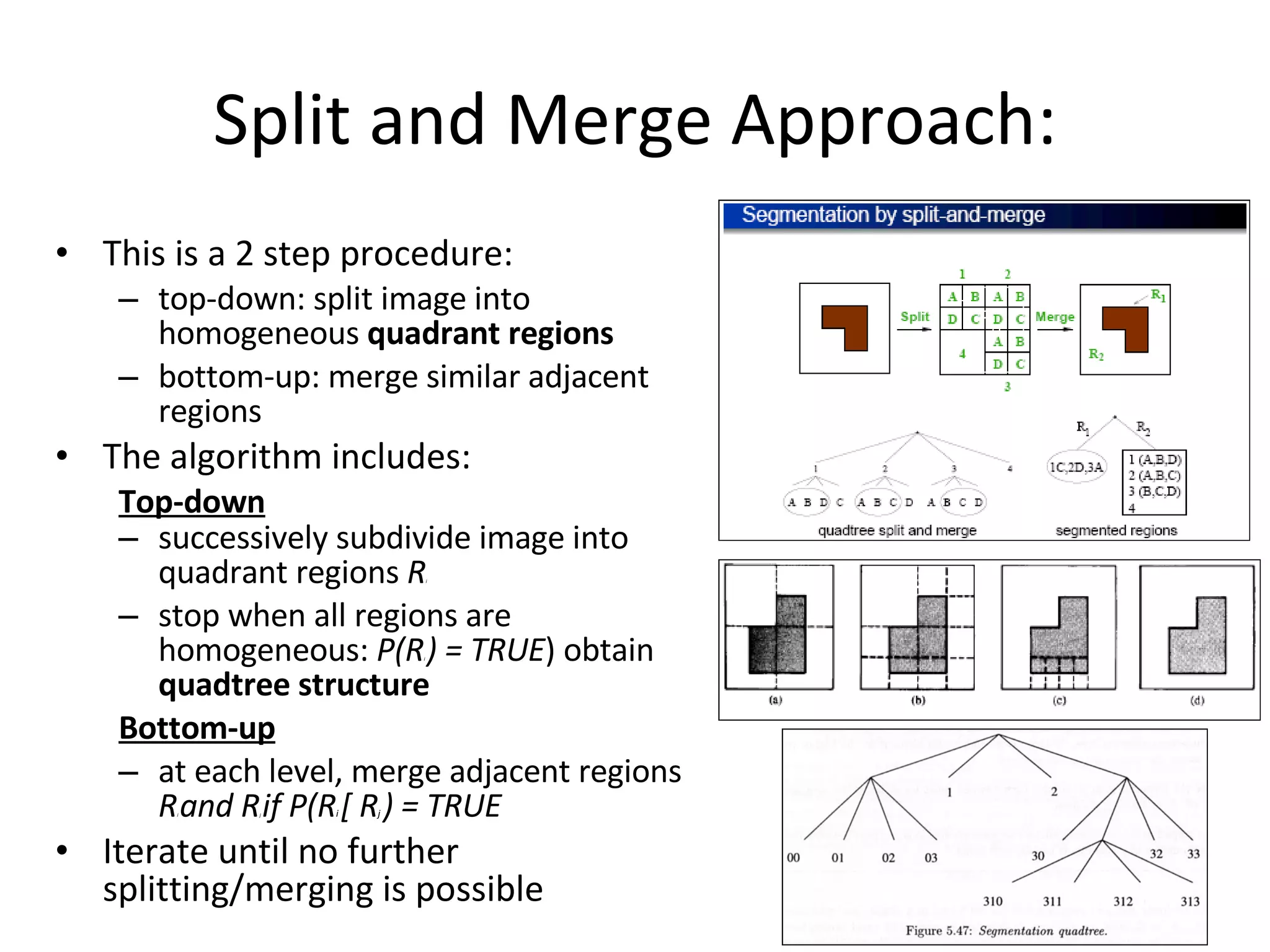
Region splitting and merging refine image sections based on characteristics, iteratively adjusting regions until they meet homogeneity.
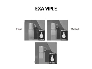
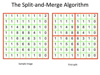
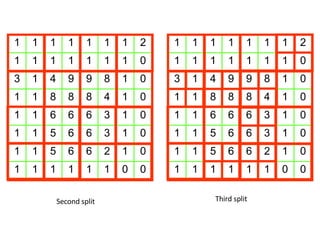
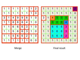
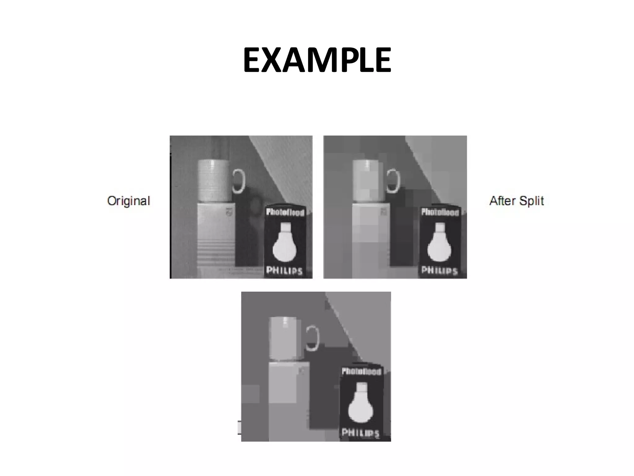
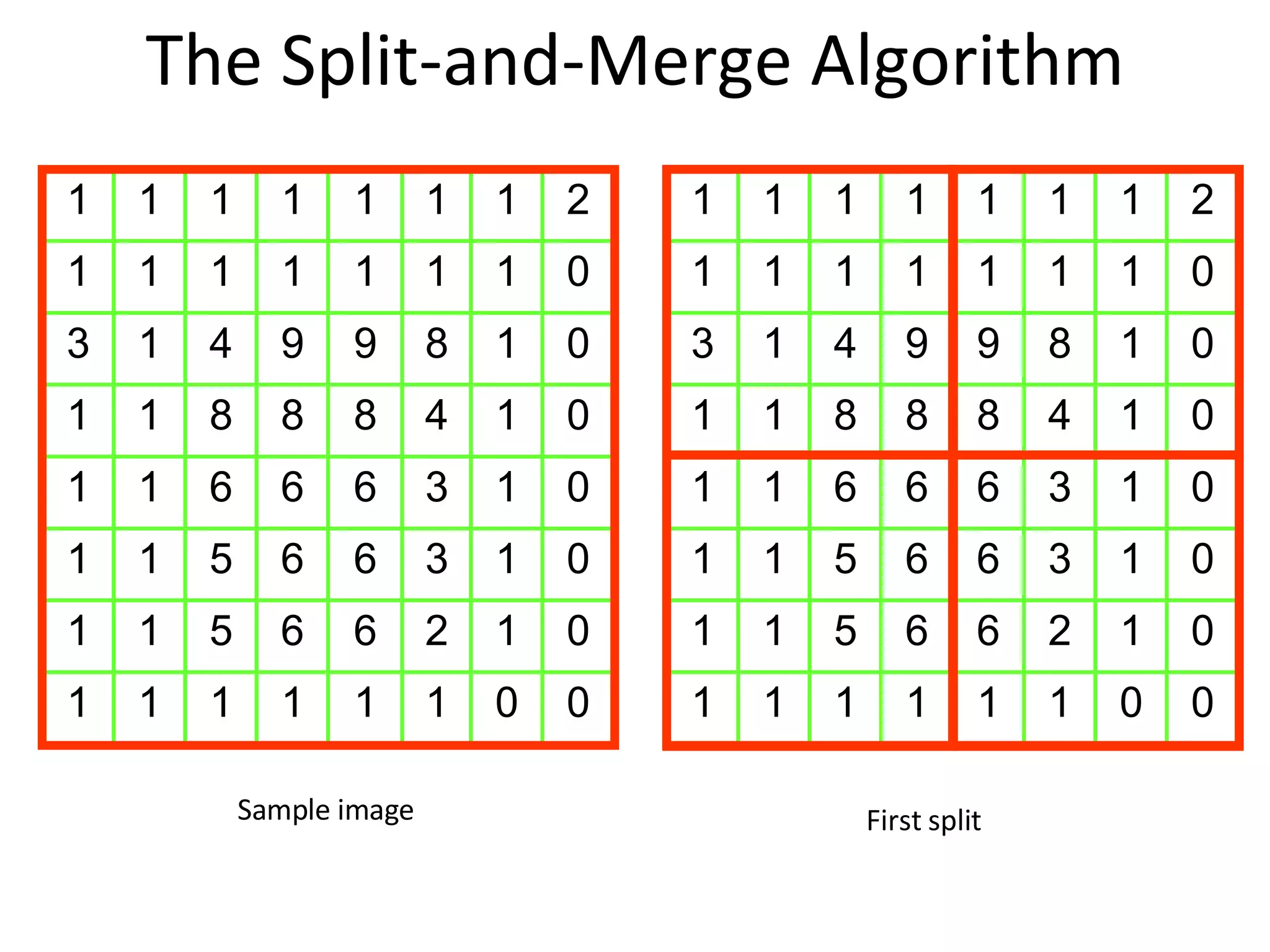
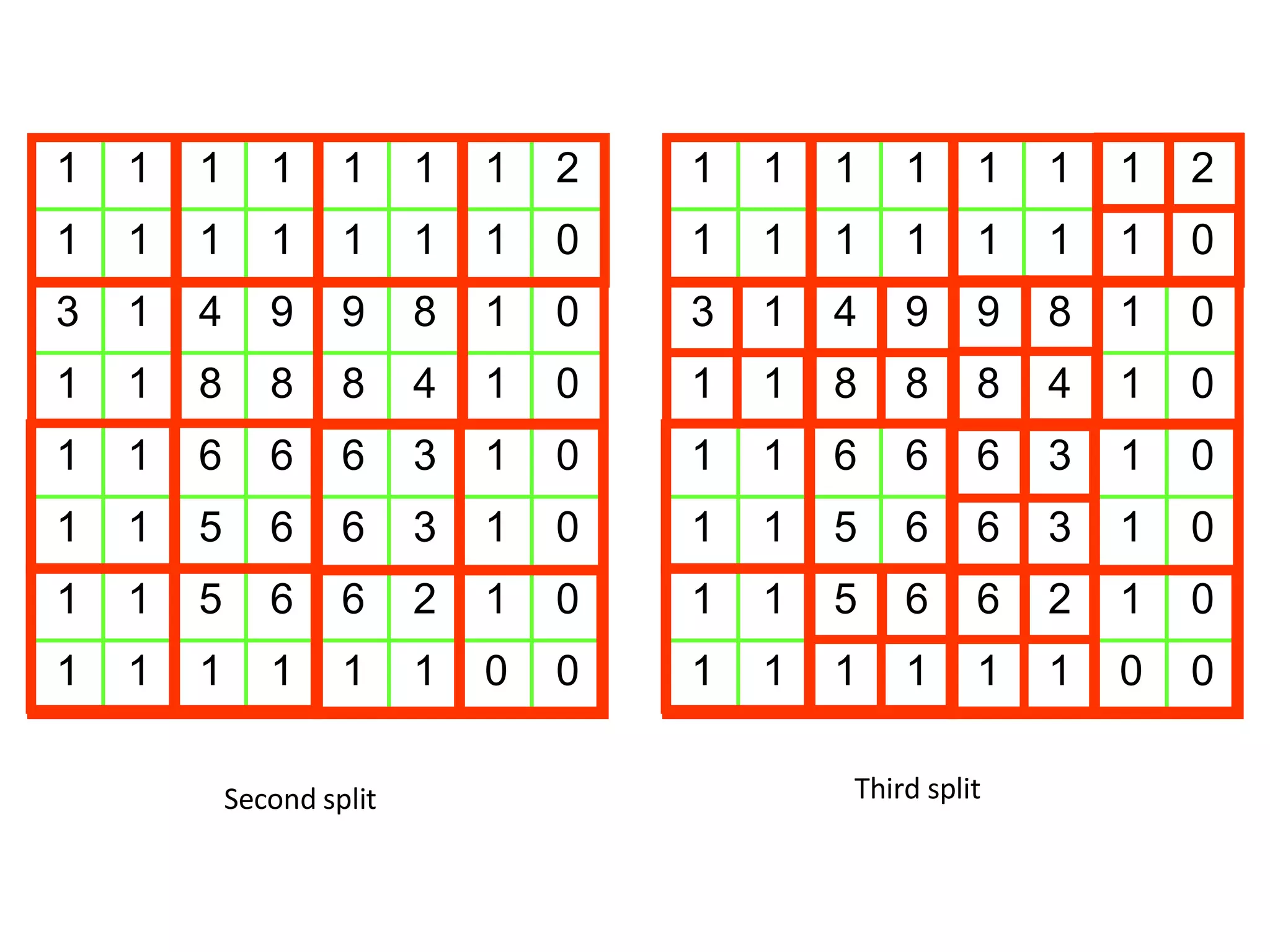
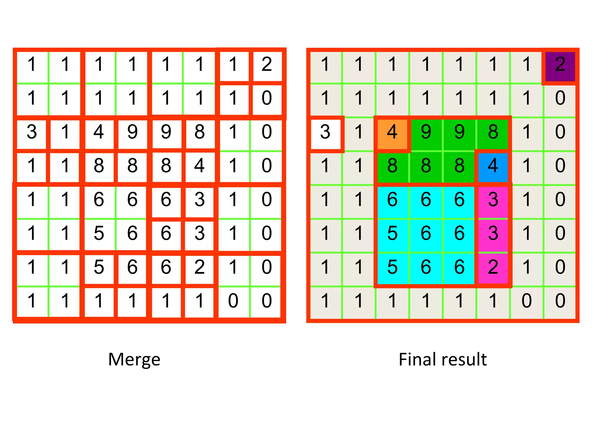
Demonstrated through sample images, the split-and-merge algorithm sequentially splits and merges regions until homogeneity is achieved.
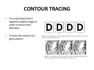
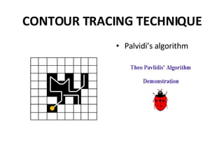
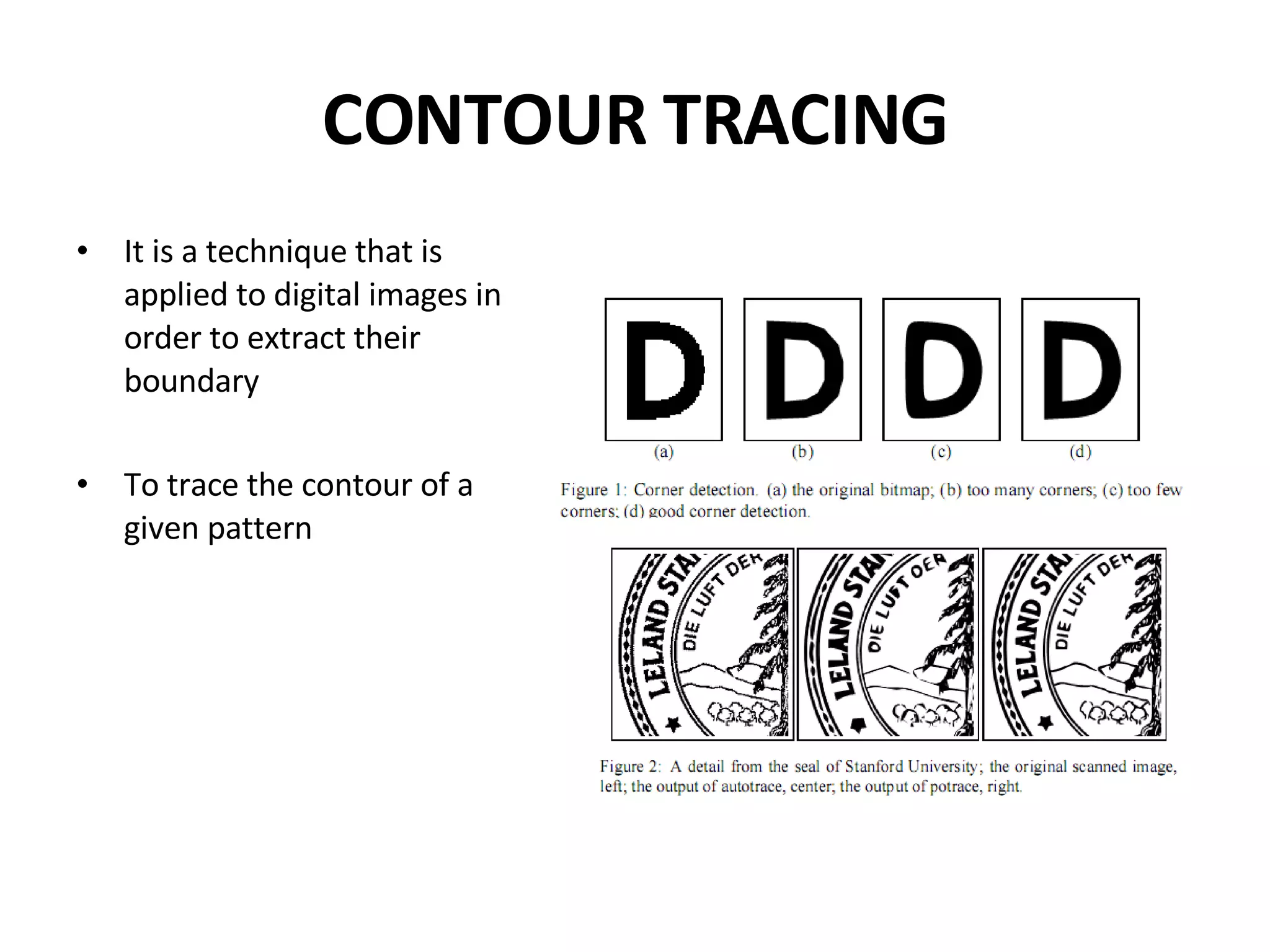
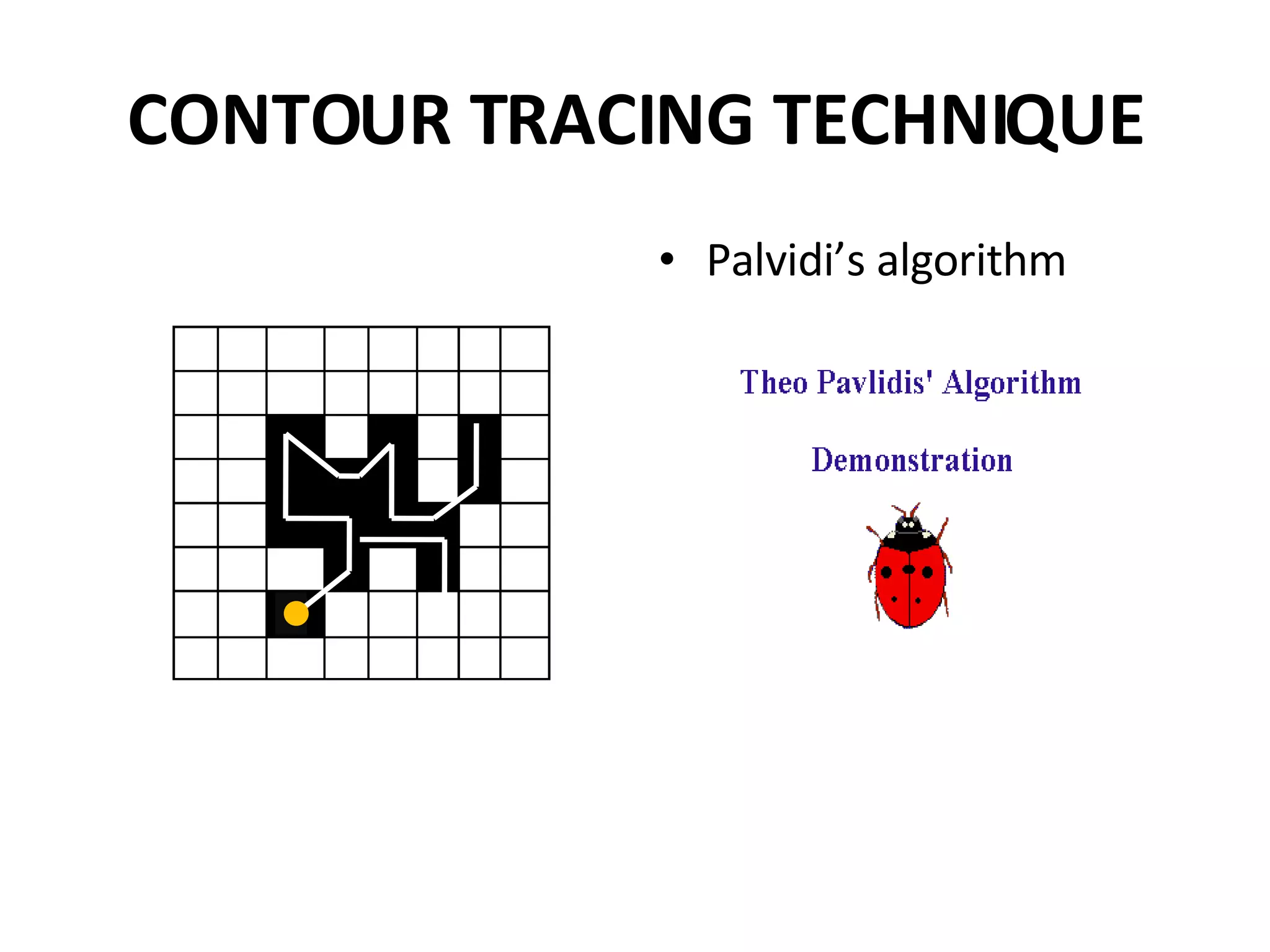
Contour tracing extracts boundaries from images using algorithms such as Palvidi's for pattern recognition.
Links provided for additional resources on edge detection and segmentation methods in image processing.
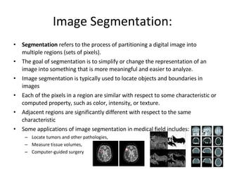
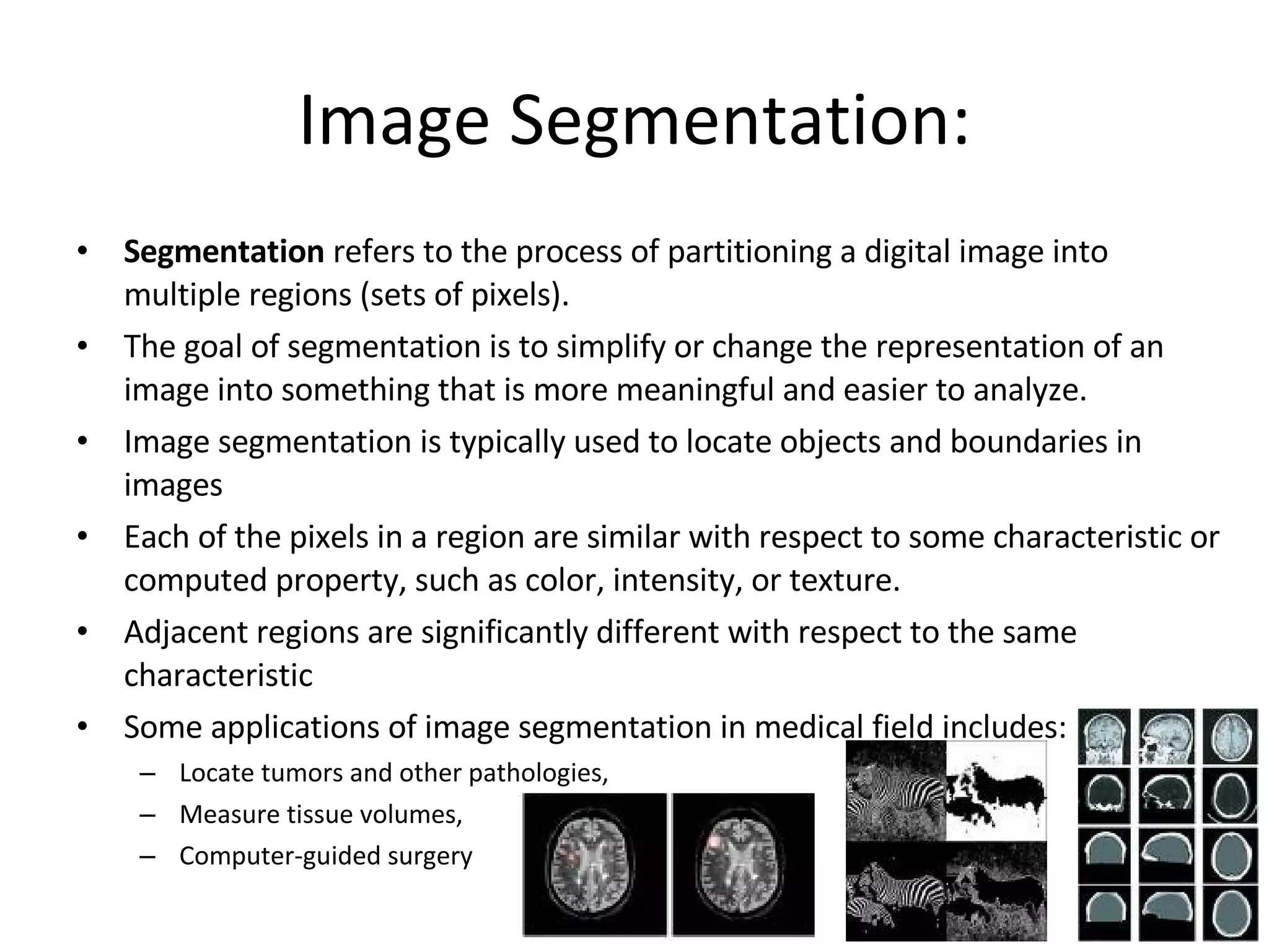
Image Segmentation: Segmentation refers to the process of partitioning a digital image into multiple regions (sets of pixels). The goal of segmentation is to simplify or change the representation of an image into something that is more meaningful and easier to analyze. Image segmentation is typically used to locate objects and boundaries in images Each of the pixels in a region are similar with respect to some characteristic or computed property, such as color, intensity, or texture. Adjacent regions are significantly different with respect to the same characteristic Some applications of image segmentation in medical field includes: Locate tumors and other pathologies, Measure tissue volumes, Computer-guided surgery
2.
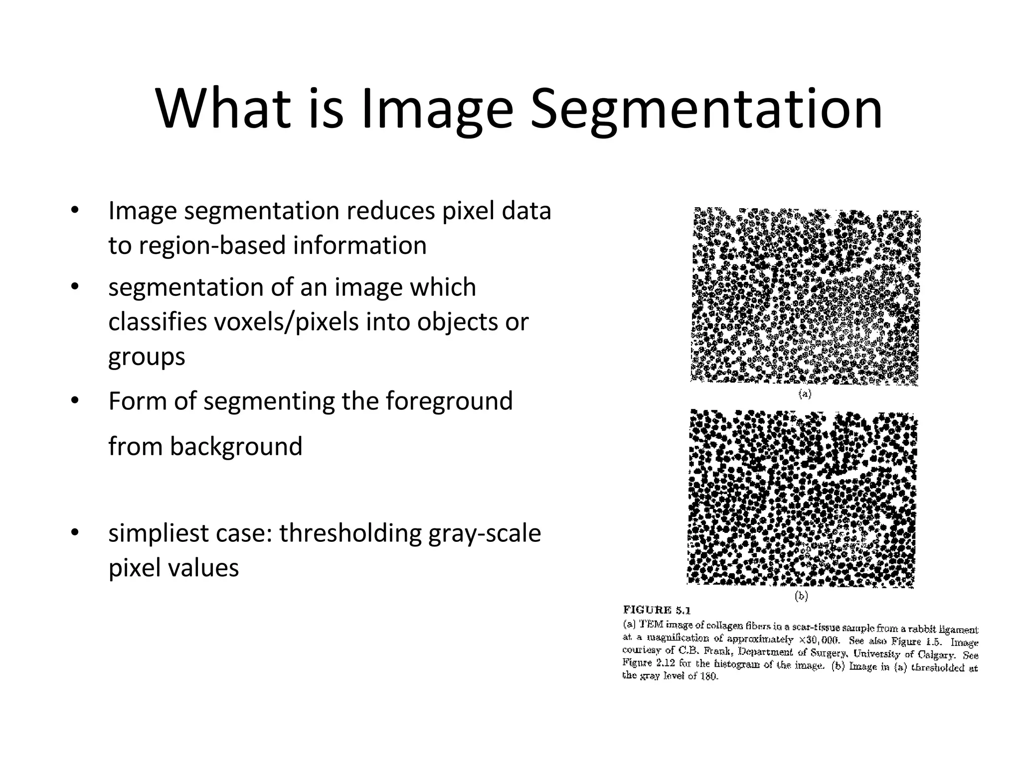
What is Image Segmentation Image segmentation reduces pixel data to region-based information segmentation of an image which classifies voxels/pixels into objects or groups Form of segmenting the foreground from background simpliest case: thresholding gray-scale pixel values
3.
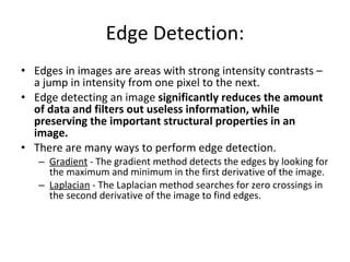
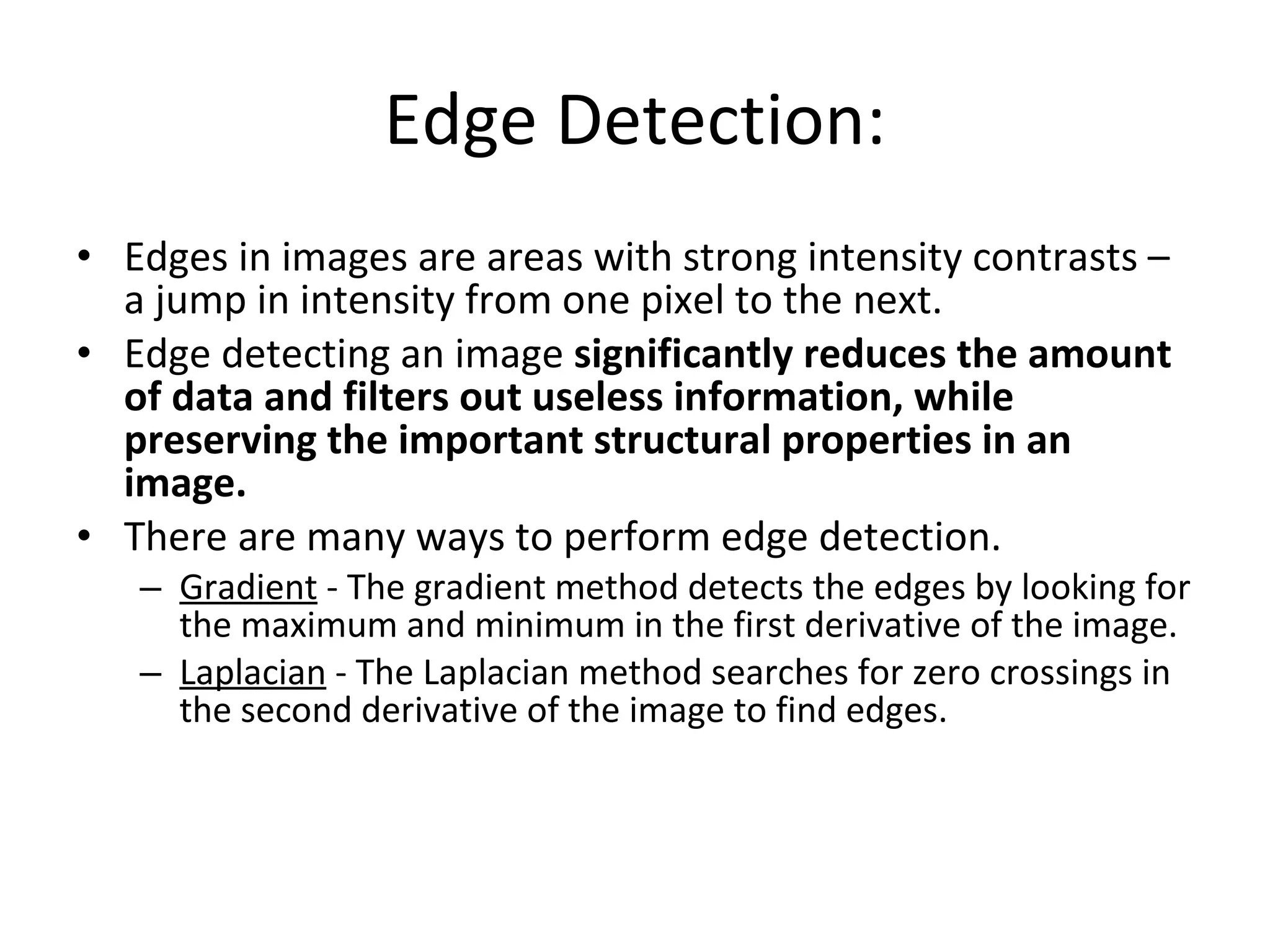
Edge Detection: Edgesin images are areas with strong intensity contrasts – a jump in intensity from one pixel to the next. Edge detecting an image significantly reduces the amount of data and filters out useless information, while preserving the important structural properties in an image. There are many ways to perform edge detection. Gradient - The gradient method detects the edges by looking for the maximum and minimum in the first derivative of the image. Laplacian - The Laplacian method searches for zero crossings in the second derivative of the image to find edges.
4.
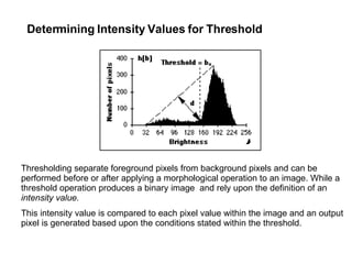
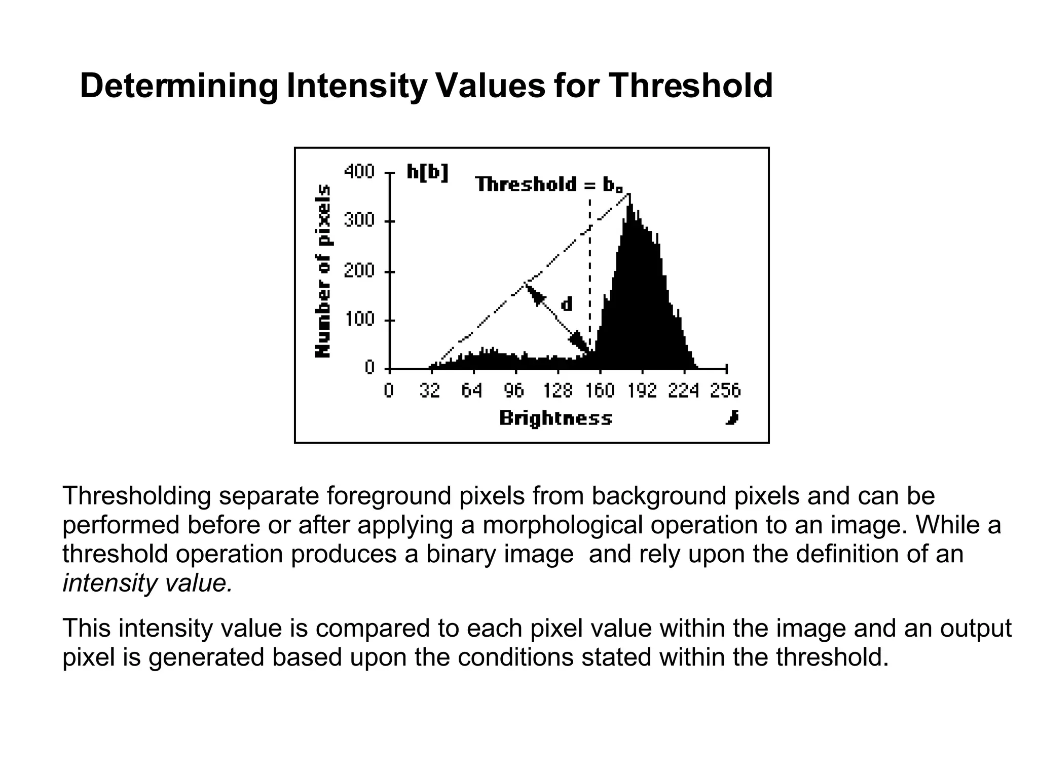
Determining Intensity Valuesfor Threshold Thresholding separate foreground pixels from background pixels and can be performed before or after applying a morphological operation to an image. While a threshold operation produces a binary image and rely upon the definition of an intensity value. This intensity value is compared to each pixel value within the image and an output pixel is generated based upon the conditions stated within the threshold.
5.
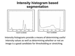
Intensity histograms providea means of determining useful intensity values as well as determining whether or not an image is a good candidate for thresholding or stretching. Intensity histogram based segmentation
6.
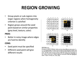
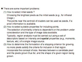
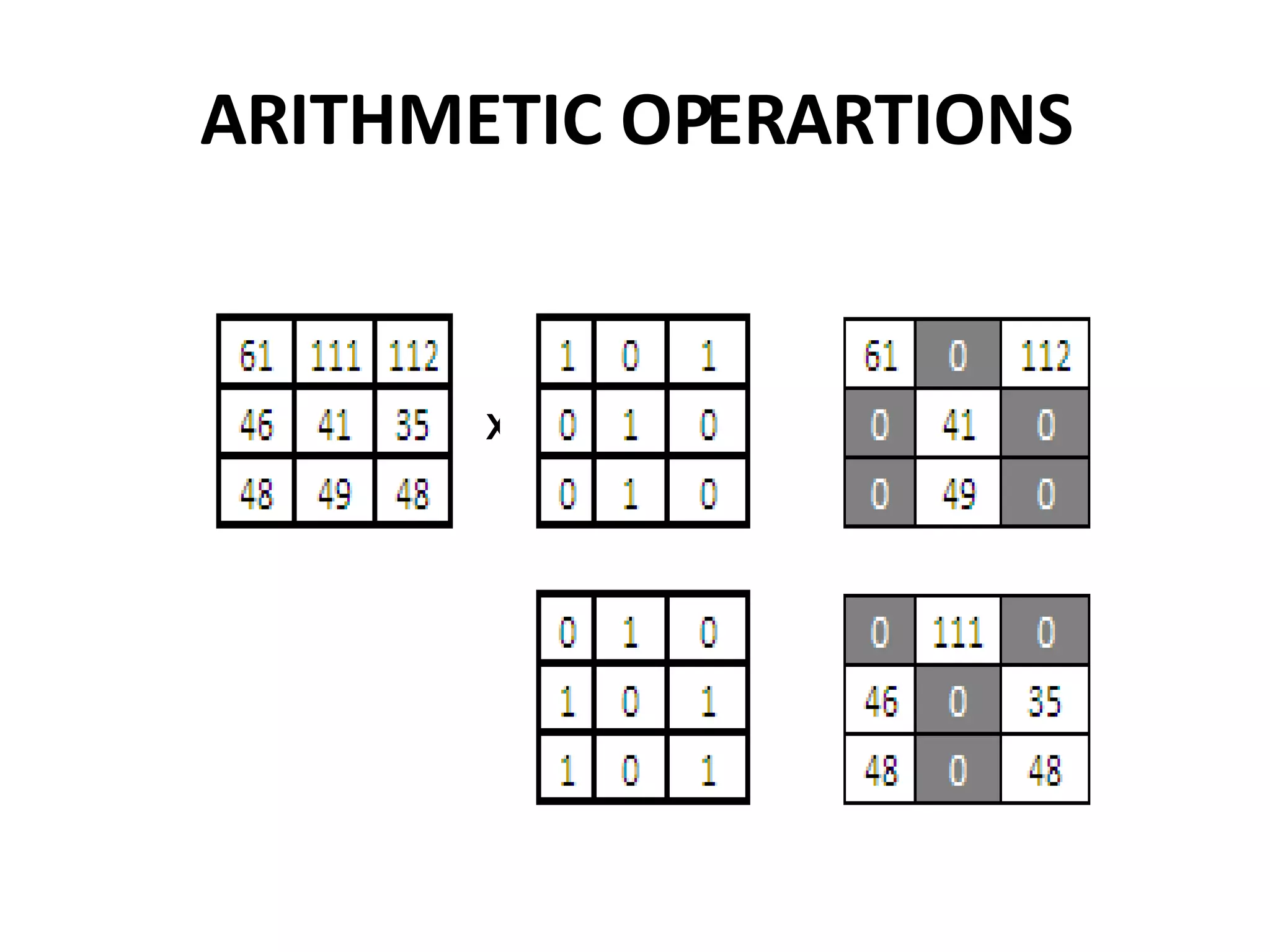
REGION GROWING Grouppixels or sub-regions into larger regions when homogeneity criterion is satisfied Region grows around the seed point based on similar properties (grey level, texture, color) PROS: Better in noisy image where edges are hard to identify CONS: Seed point must be specified Different seed point will give different results PIXEL AGGREGATION: Homogeneity criteria: The difference between 2 pixel values is less than or equal to 5 Horizontal, vertical, diagonal 10 10 10 10 10 10 10 10 10 10 69 70 10 10 59 10 60 64 59 56 60 10 59 10 60 70 10 62 10 60 59 65 67 10 65 10 10 10 10 10 10 10 10 10 10 10 10 10 10
7.
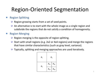
Region-Oriented Segmentation RegionSplitting Region growing starts from a set of seed points. An alternative is to start with the whole image as a single region and subdivide the regions that do not satisfy a condition of homogeneity. Region Merging Region merging is the opposite of region splitting. Start with small regions (e.g. 2x2 or 4x4 regions) and merge the regions that have similar characteristics (such as gray level, variance). Typically, splitting and merging approaches are used iteratively.
8.
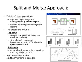
Split and MergeApproach: This is a 2 step procedure: top-down: split image into homogeneous quadrant regions bottom-up: merge similar adjacent regions The algorithm includes: Top-down successively subdivide image into quadrant regions R i stop when all regions are homogeneous: P(R i ) = TRUE ) obtain quadtree structure Bottom-up at each level, merge adjacent regions R i and R j if P(R i [ R j ) = TRUE Iterate until no further splitting/merging is possible
#2 The result of image segmentation is a set of regions that collectively cover the entire image, or a set of contours extracted from the image (see edge detection ). Each of the pixels in a region are similar with respect to some characteristic or computed property, such as color , intensity , or texture . Adjacent regions are significantly different with respect to the same characteristic(s)