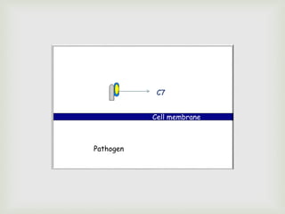
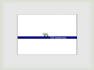
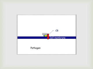
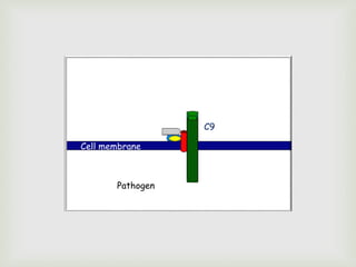
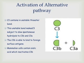
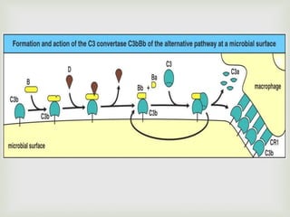
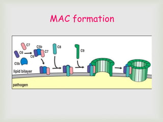
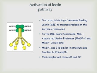
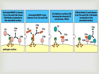
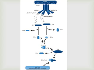
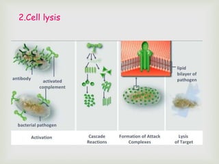
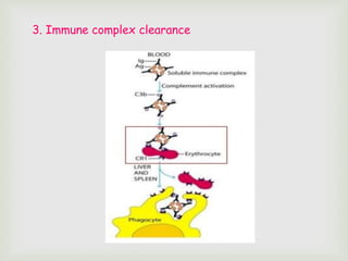
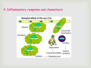
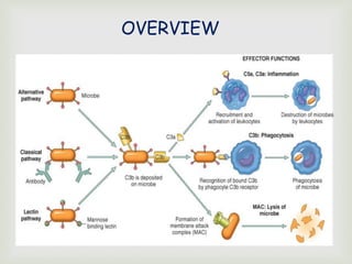
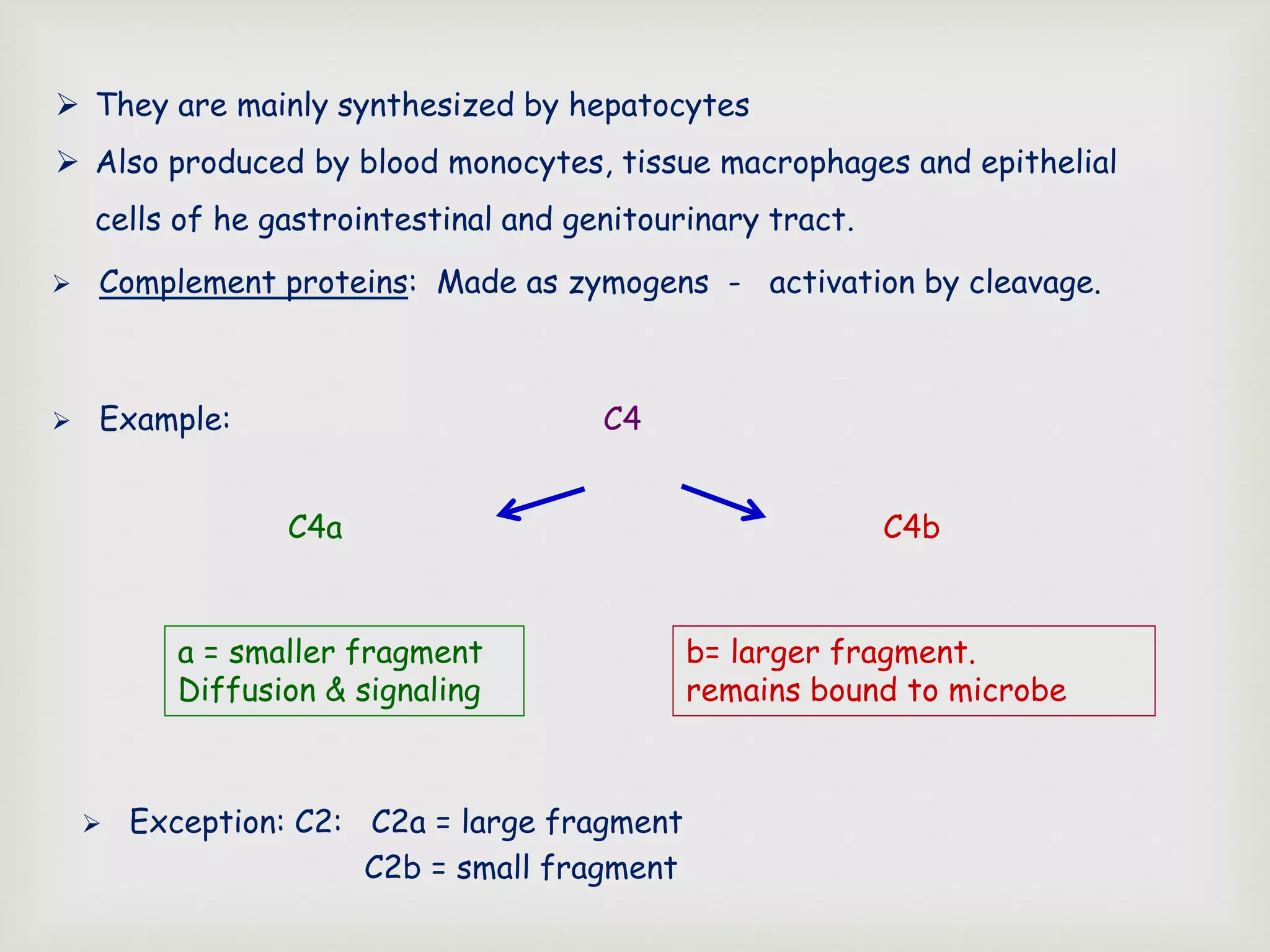
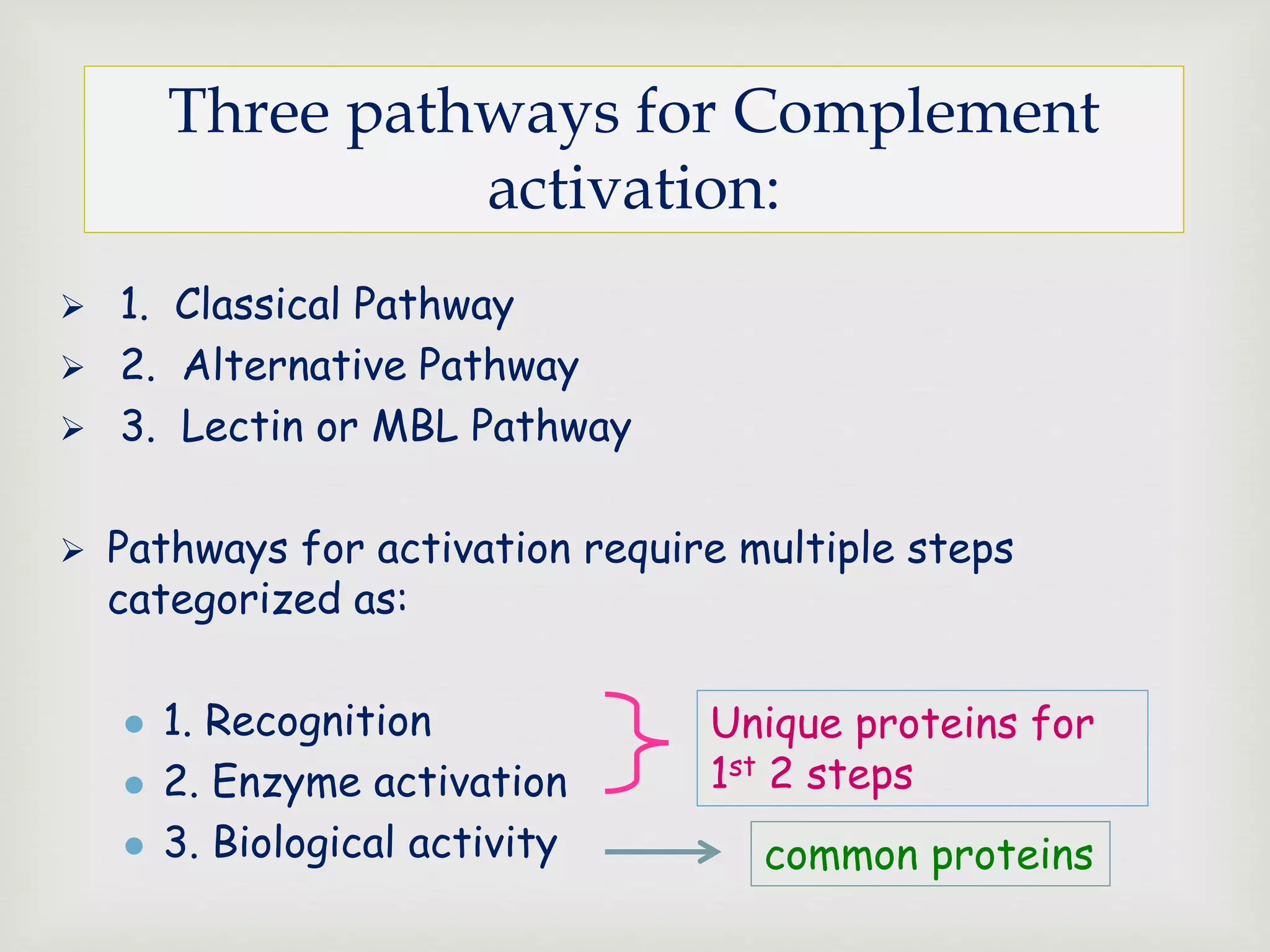
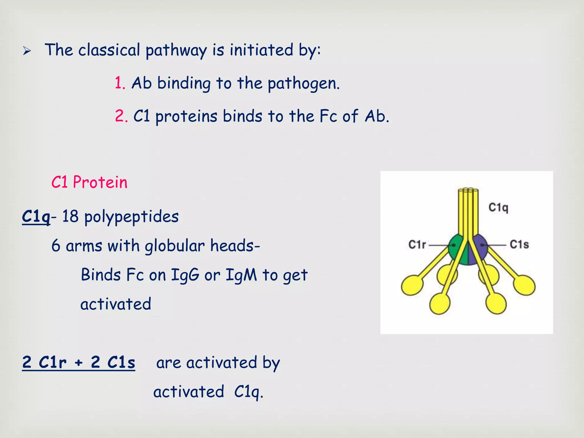
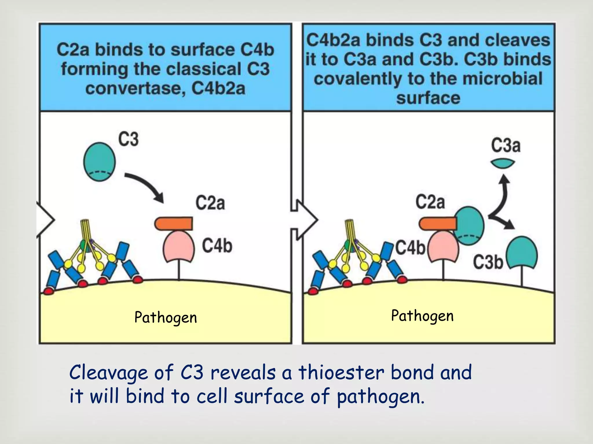
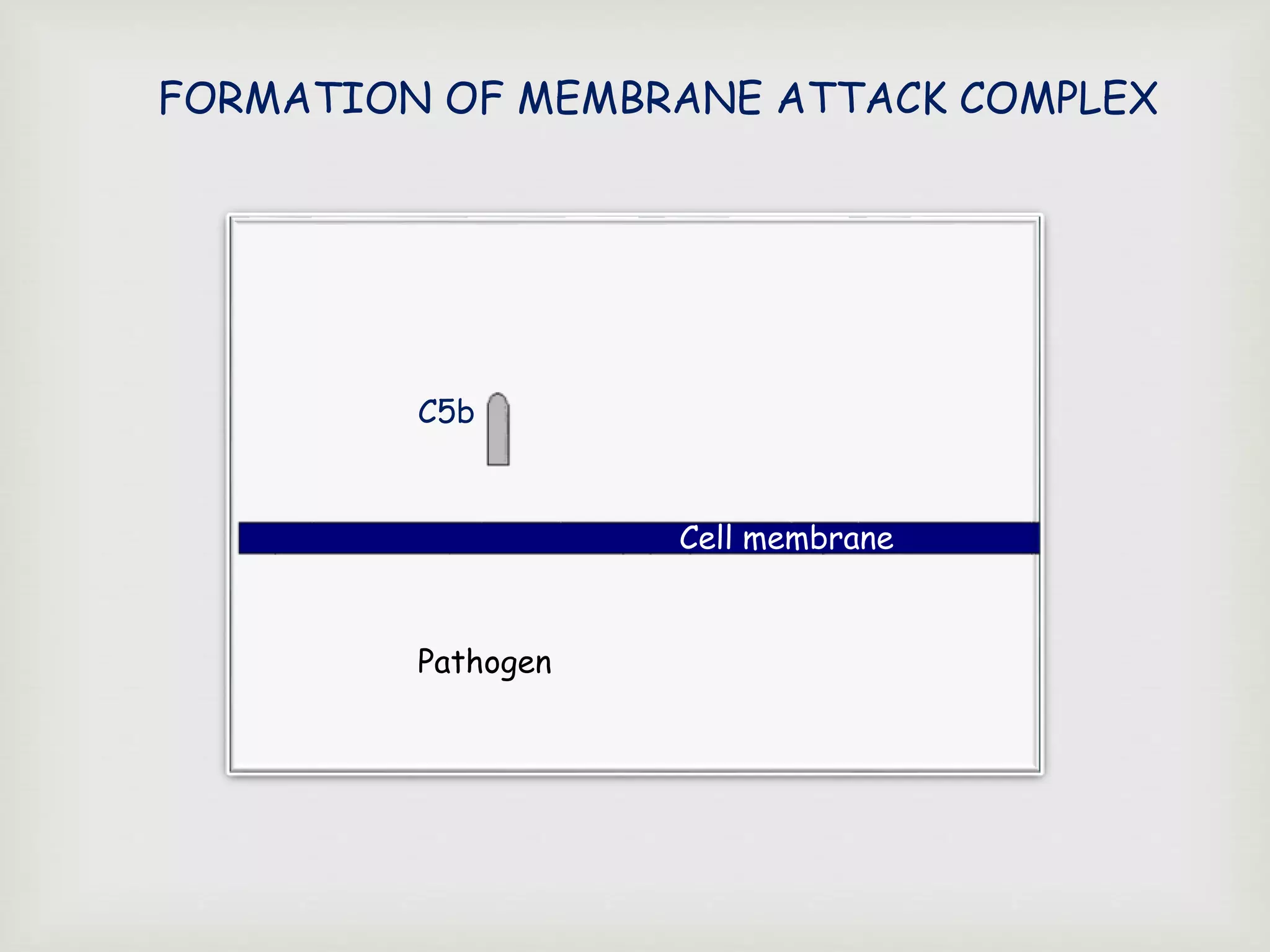
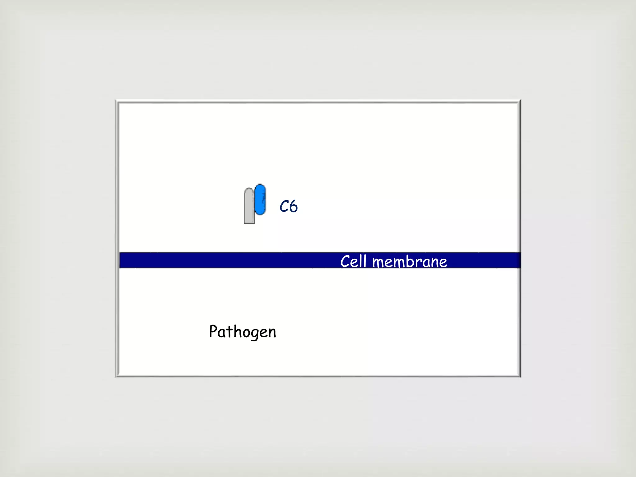
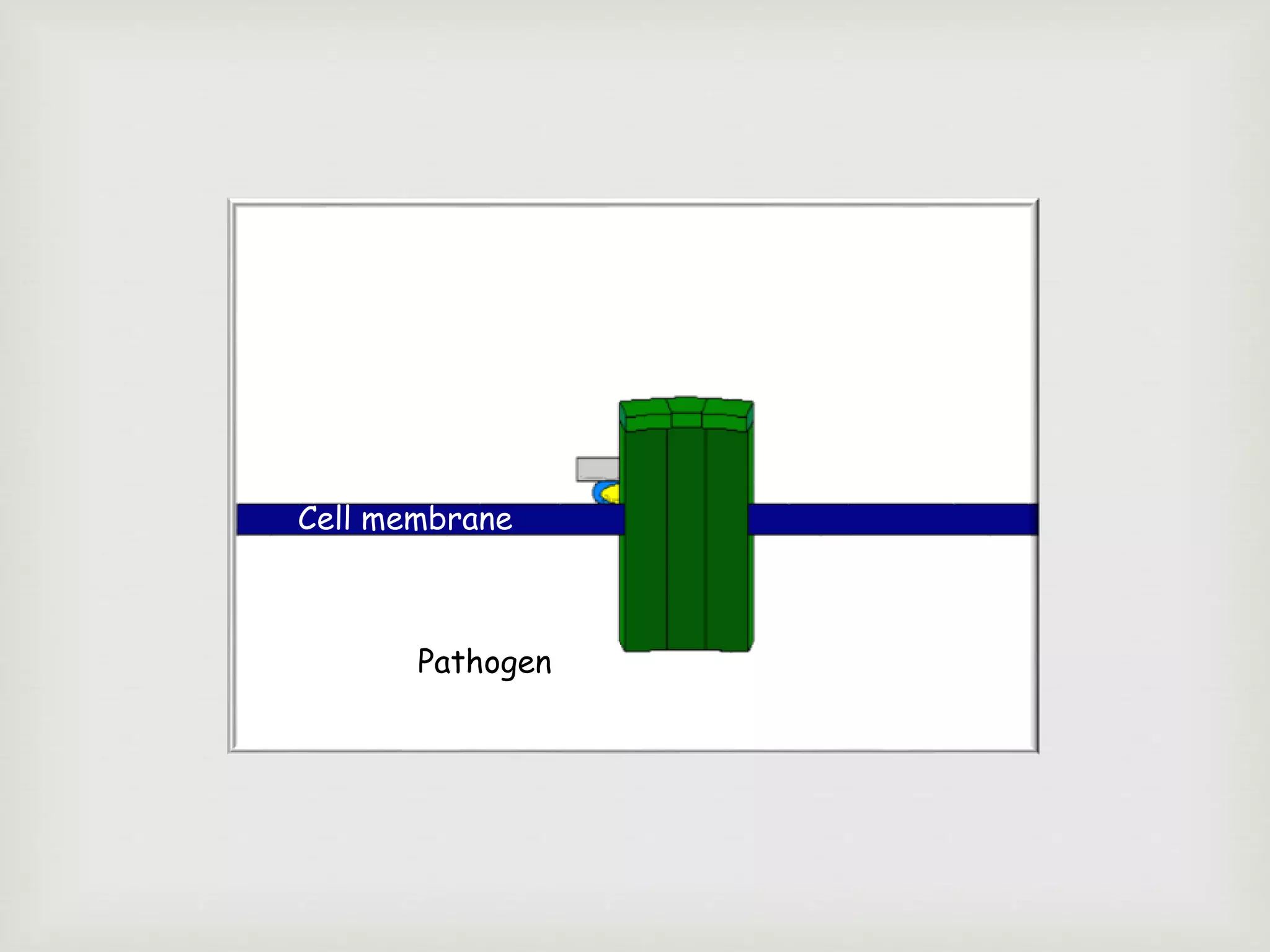
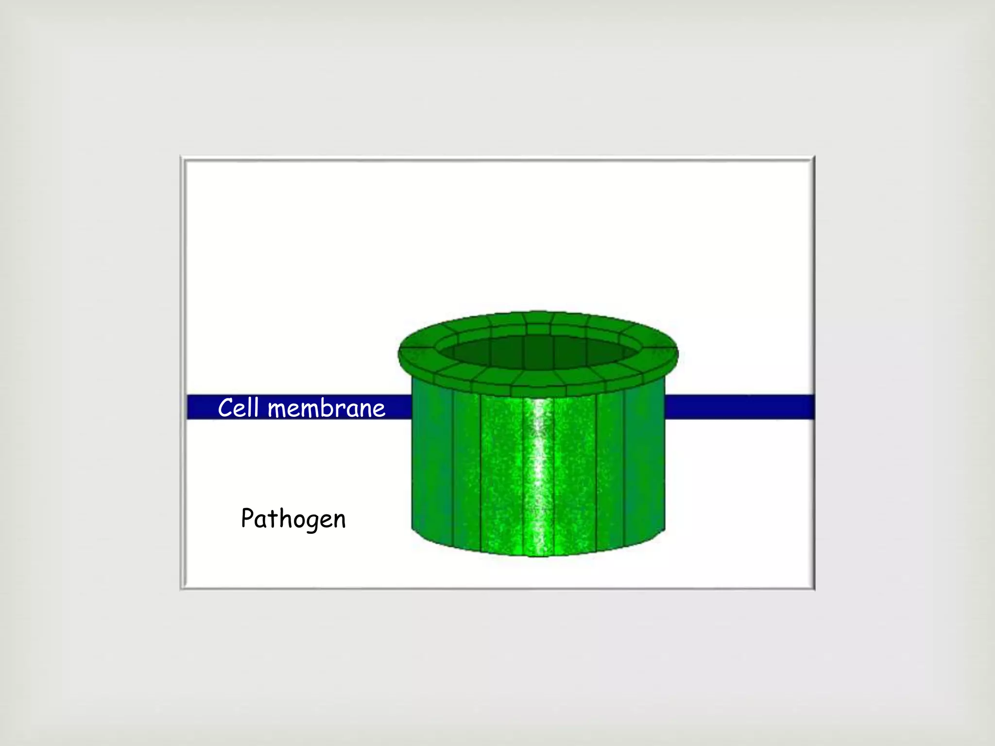
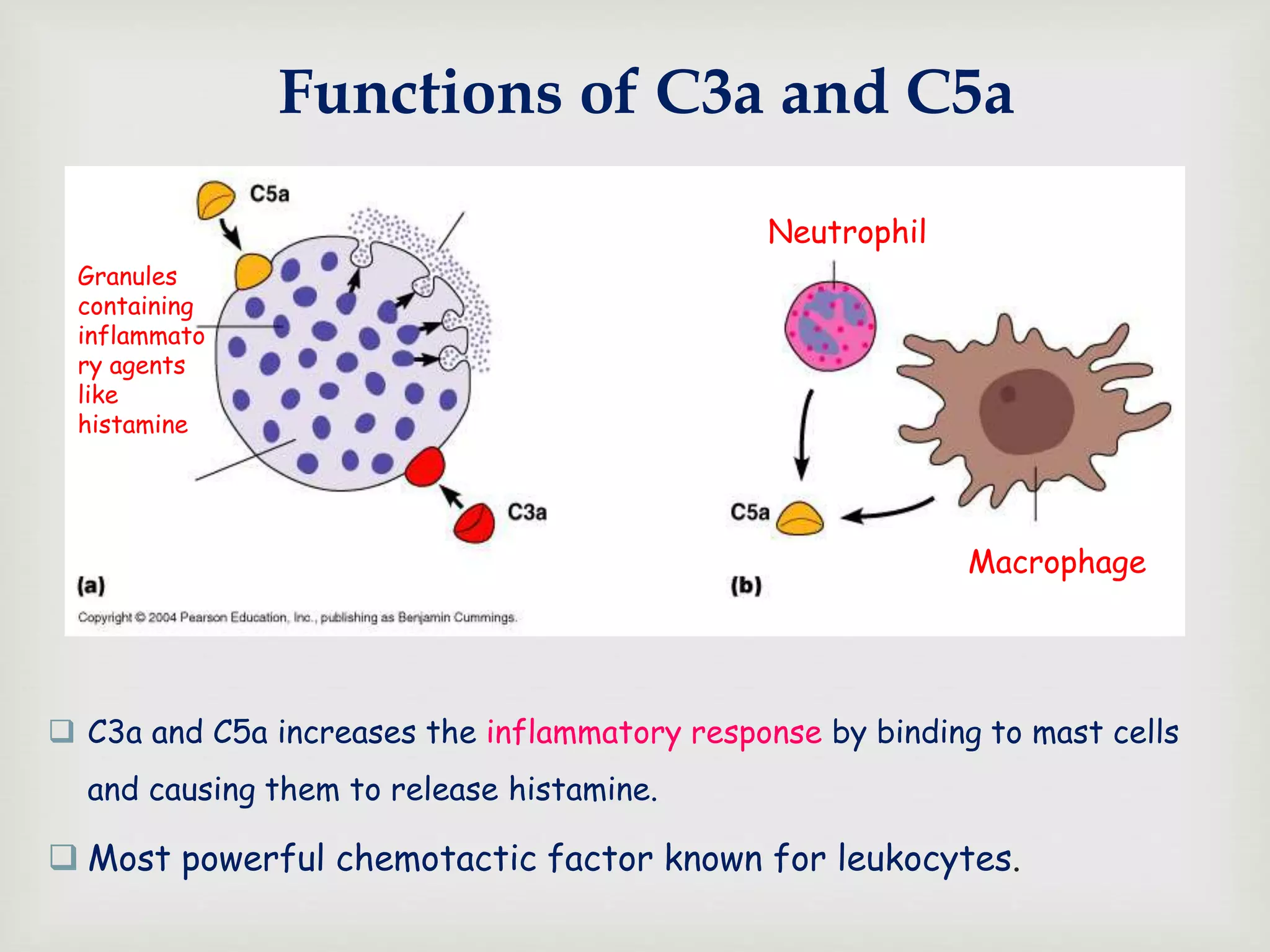
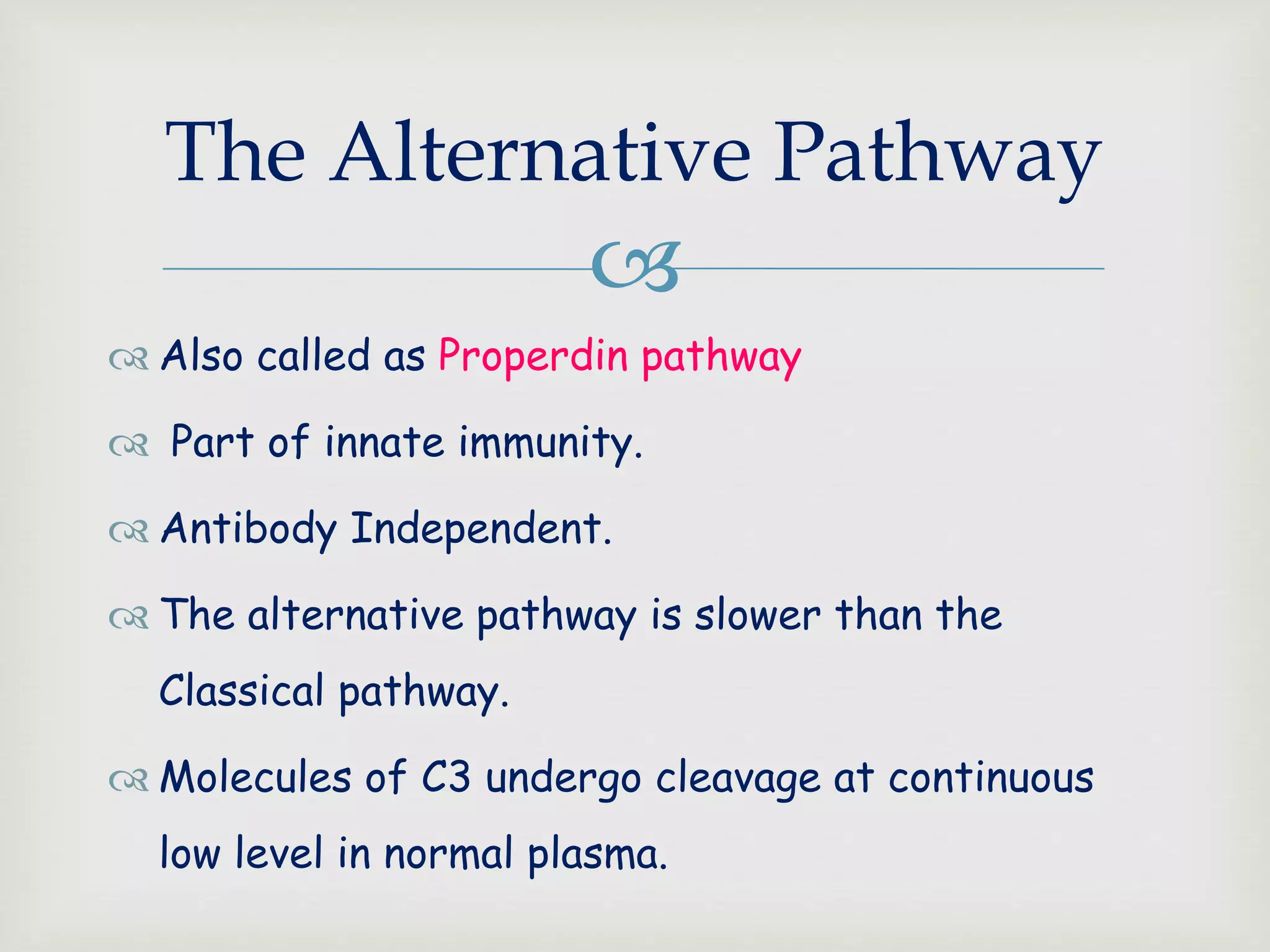
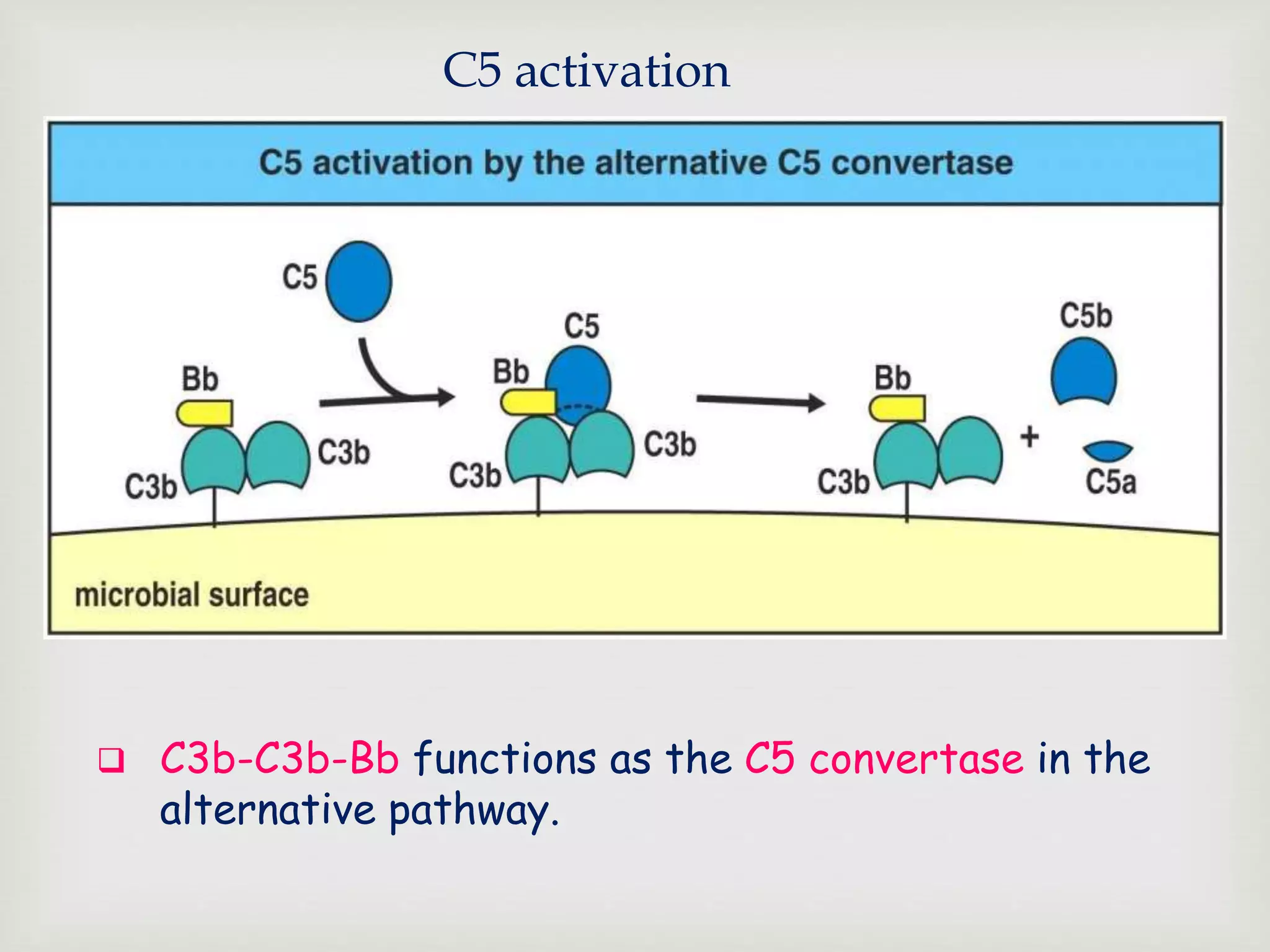
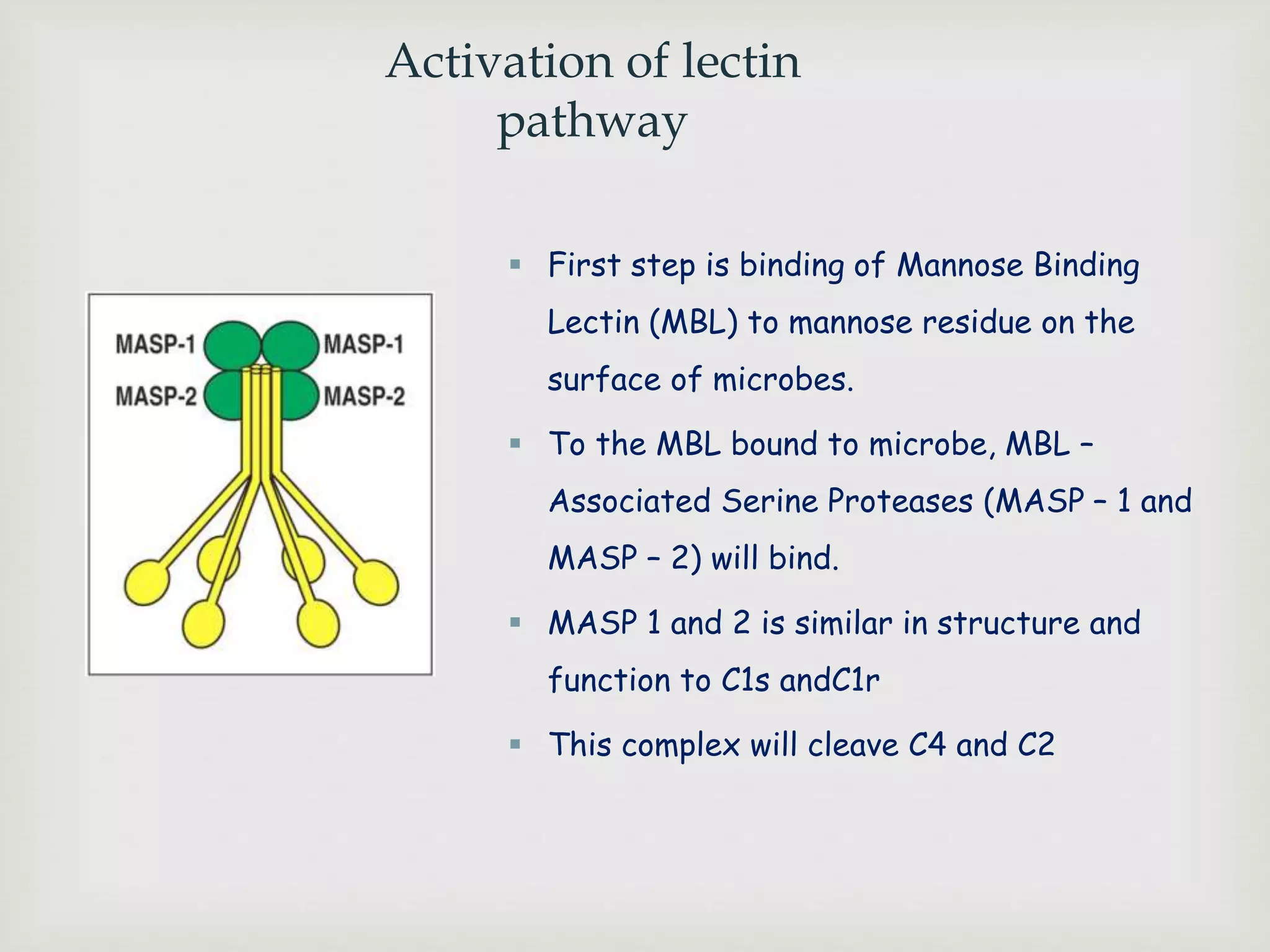
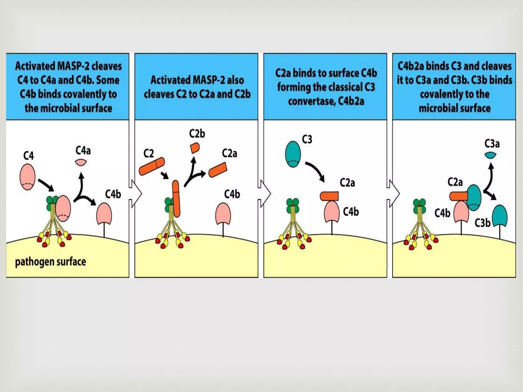
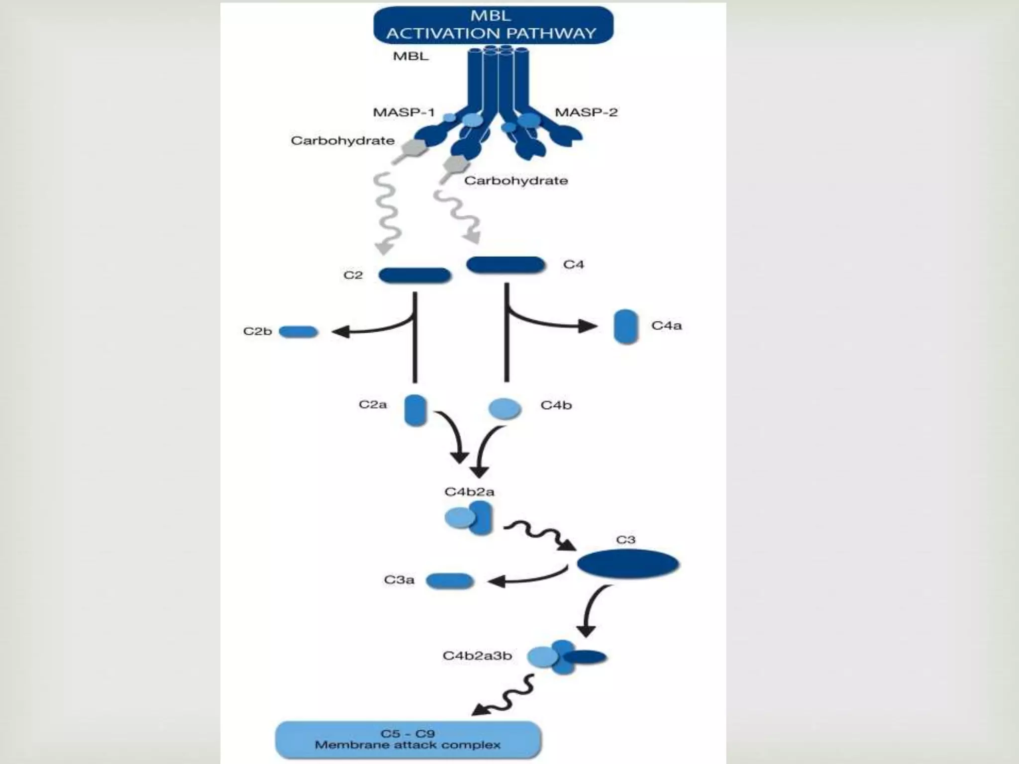
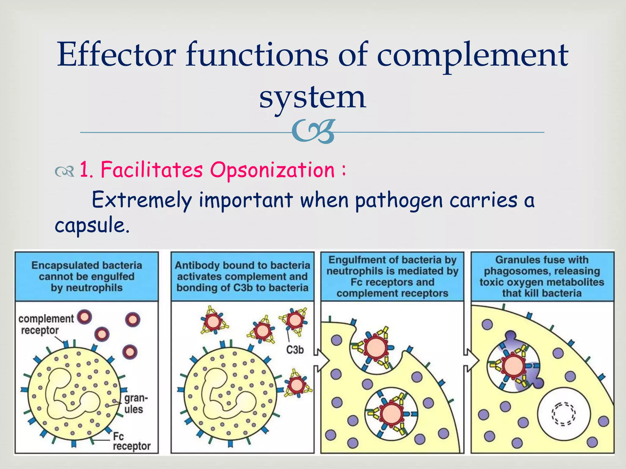
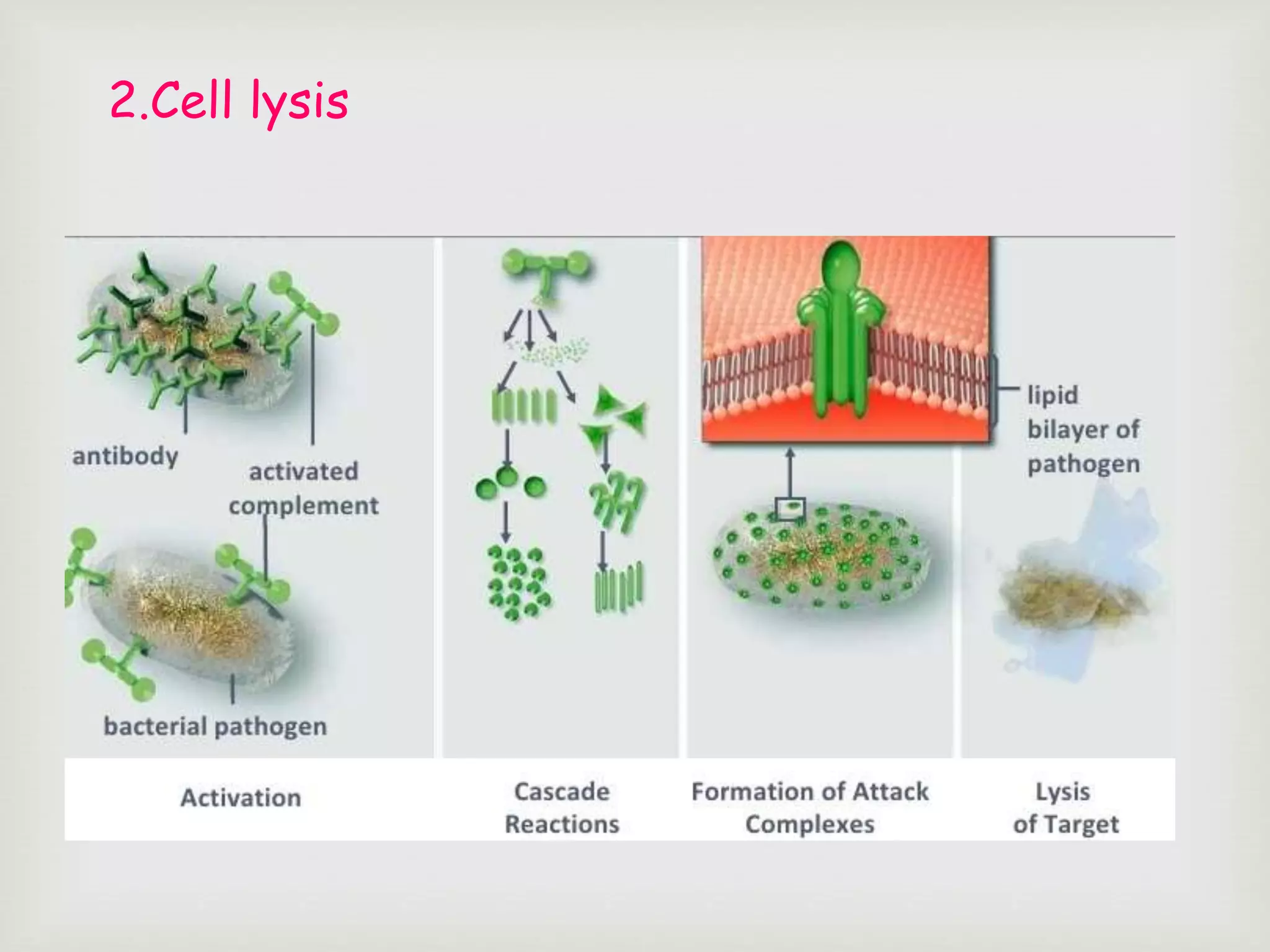
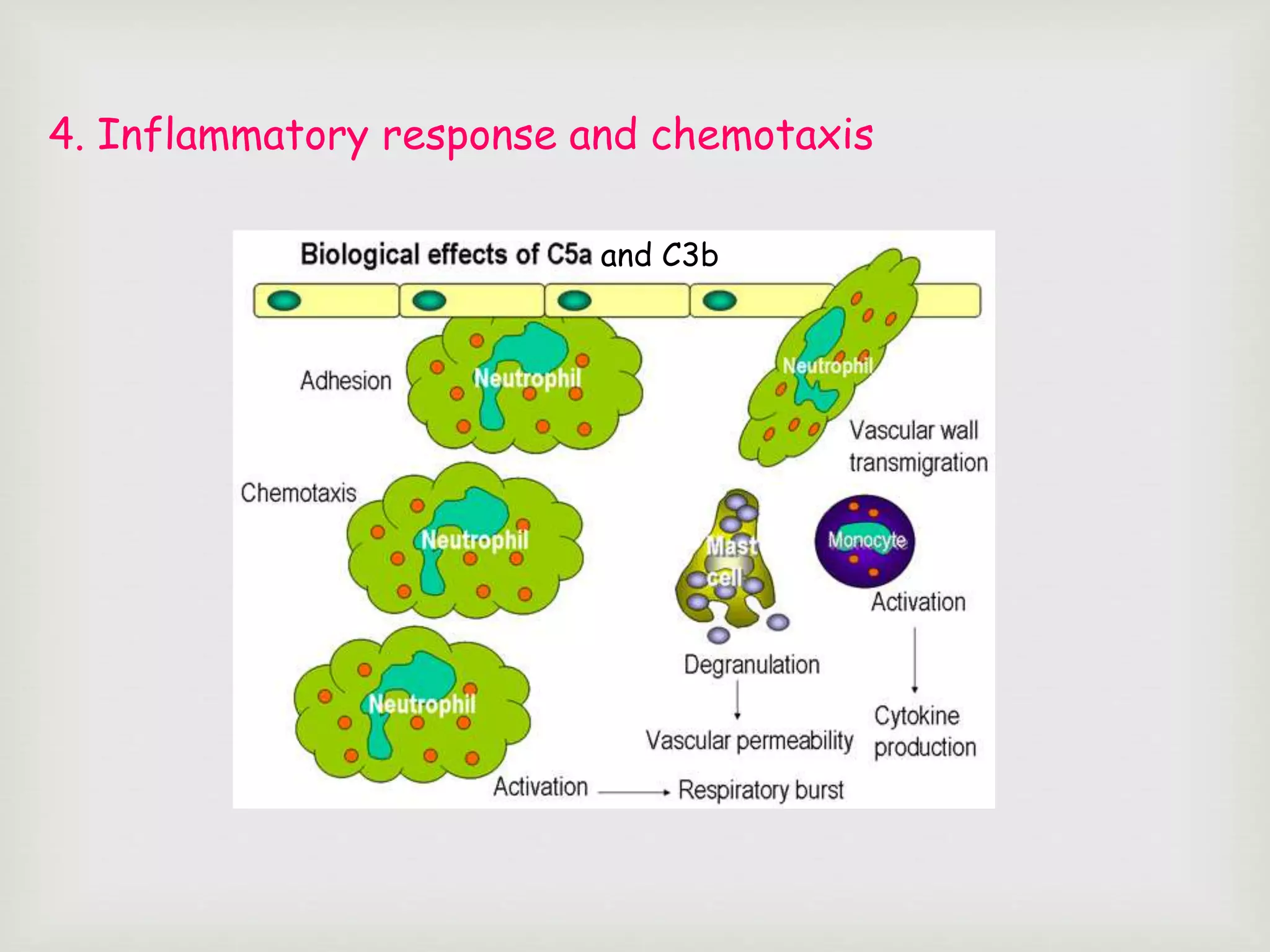
KEMBAR78
The complement system | PPTX
PRESENTED BY :
Sushma P.R
M.Sc. Biotech 2nd sem
Dept. of biotechnology
PRESENTED TO :
Dr. T.T.S Ramachandra Murthy
HOD, Dept. of Biotechnology
Brindavan college

 Research in complement started in 1890s when Jules Bordet
at the Institut Pasteur of Paris conducted experiment using
sheep antiserum.
 He named those substances as
Alexins.
 Paul Ehrlich coined the term
complement.
HISTORY

 It is named “complement system” because it was first identified as
a heat-labile component of serum that “complemented” antibodies
in the killing of bacteria.
 Consists of serum and cell surface proteins involved in defense
against pathogens and tissue damage mediated by antibodies
 The Complement system is the major effector of humoral branch
of immune system.
 Plays major role in both innate and adaptive immunity.
INTRODUCTION

 Opsonisation
 Chemotaxis
 Cell lysis
 Immune clearance
 Activation of inflammatory response
Functions of the complement
system

 Over 30 serum and cell surface proteins:
- Complement components
(in serum inactive, activated sequentially as a cascade)
- Complement receptors
(cell surface, recognize activated components)
- Regulatory proteins of complement
(both in serum and cell surface, inhibit activated components)
Components are designated by numbers (E.g. ; C1 – C9) or latters
(E.g. : Factor D).
The components of complement
system
 Complement proteins: Made as zymogens - activation by cleavage.
 Example: C4
 Exception: C2: C2a = large fragment
C2b = small fragment
a = smaller fragment
Diffusion & signaling
b= larger fragment.
remains bound to microbe
C4a C4b
 They are mainly synthesized by hepatocytes
 Also produced by blood monocytes, tissue macrophages and epithelial
cells of he gastrointestinal and genitourinary tract.
Three pathways for Complement
activation:
 1. Classical Pathway
 2. Alternative Pathway
 3. Lectin or MBL Pathway
 Pathways for activation require multiple steps
categorized as:
 1. Recognition
 2. Enzyme activation
 3. Biological activity
Unique proteins for
1st 2 steps
common proteins
THE CLASSICAL PATHWAY
 Part of adaptive immune system
 Relies on Ag – Ab complex to get activated
 The classical pathway is initiated by:
1. Ab binding to the pathogen.
2. C1 proteins binds to the Fc of Ab.
C1 Protein
C1q- 18 polypeptides
6 arms with globular heads-
Binds Fc on IgG or IgM to get
activated
2 C1r + 2 C1s are activated by
activated C1q.
Planar structure of
IgM
Doesn’t have any exposed
C1q binding site.
Staple structure
of IgM
At least 3 binding sited
for C1q are exposed.
Cleavage of C3 reveals a thioester bond and
it will bind to cell surface of pathogen.
Pathogen Pathogen
C3b
C3b is an opsonin
Opsonins are molecules
that bind both to bacteria
and phagocytes
Opsonization increases
phagocytosis by 1,000 fold.
C3b s attached to
microbial surface
microbe
 C4b-2a-3b functions as the classical
C5 convertase:
FORMATION OF MEMBRANE ATTACK COMPLEX
C5b
Cell membrane
Pathogen
C6
Cell membrane
Pathogen
Cell membrane
Pathogen
C7
Cell membrane
Cell membrane
Pathogen
C8
Cell membrane
Pathogen
C9
Cell membrane
Pathogen
Cell membrane
Pathogen
Cell membrane
Pathogen
Cell membrane
Pathogen
Cell membrane
Pathogen
Cell membrane
Pathogen
Functions of C3a and C5a
 C3a and C5a increases the inflammatory response by binding to mast cells
and causing them to release histamine.
 Most powerful chemotactic factor known for leukocytes.
Neutrophil
Macrophage
Granules
containing
inflammato
ry agents
like
histamine

The Alternative Pathway
 Also called as Properdin pathway
 Part of innate immunity.
 Antibody Independent.
 The alternative pathway is slower than the
Classical pathway.
 Molecules of C3 undergo cleavage at continuous
low level in normal plasma.
Activation of Alternative
pathway
 C3 contains in unstable thioester
bond.
 This unstable bond makesC3
subject to slow spontaneous
hydrolysis to C3b and C3a
 The C3b is able to bind to foreign
surface antigens.
 Mammalian cells contain sialic
acid which inactivates C3b
Factor B
 C3b on the surface of a
foreign cells binds to
another plasma protein
called factor B
 This binding of Factor B
results in the exposure
of a binding site for
another enzyme called
Factor D
Factor D
 The binding of factor B to
C3b allows a protein
enzyme called Factor D to
cleave Factor B to Ba and
Bb.
 Factor Bb remains bound
to C3b while Ba and Factor
D disperse away.
 The complex C3bBb is
C3 convertase of
alternative pathway
Ba
Properdin- Factor P
 Stabilizes C3b-Bb so it may cleave more C3.
May cleave over a million C3 molecules!
 Protects Bb-C3b from inactivation by
complement control mechanisms.
C5 activation
 C3b-C3b-Bb functions as the C5 convertase in the
alternative pathway.
MAC formation

 Independent of antibodies.
 Lectins are proteins that bind to specific cb targets.
 Activated by Mannose binding lectin (MBL) – lectin that
binds to mannose residues on the microbes.
 MBL is similar to C1q in structure and function.
Lectin Binding Pathway
Activation of lectin
pathway
 First step is binding of Mannose Binding
Lectin (MBL) to mannose residue on the
surface of microbes.
 To the MBL bound to microbe, MBL –
Associated Serine Proteases (MASP – 1 and
MASP – 2) will bind.
 MASP 1 and 2 is similar in structure and
function to C1s andC1r
 This complex will cleave C4 and C2

 1. Facilitates Opsonization :
Extremely important when pathogen carries a
capsule.
Effector functions of complement
system
2.Cell lysis
3. Immune complex clearance
and C3b
4. Inflammatory response and chemotaxis
OVERVIEW
THANK YOU

The complement system

  • 1.
    PRESENTED BY : SushmaP.R M.Sc. Biotech 2nd sem Dept. of biotechnology PRESENTED TO : Dr. T.T.S Ramachandra Murthy HOD, Dept. of Biotechnology Brindavan college
  • 2.
      Research incomplement started in 1890s when Jules Bordet at the Institut Pasteur of Paris conducted experiment using sheep antiserum.  He named those substances as Alexins.  Paul Ehrlich coined the term complement. HISTORY
  • 3.
      It isnamed “complement system” because it was first identified as a heat-labile component of serum that “complemented” antibodies in the killing of bacteria.  Consists of serum and cell surface proteins involved in defense against pathogens and tissue damage mediated by antibodies  The Complement system is the major effector of humoral branch of immune system.  Plays major role in both innate and adaptive immunity. INTRODUCTION
  • 4.
      Opsonisation  Chemotaxis Cell lysis  Immune clearance  Activation of inflammatory response Functions of the complement system
  • 6.
      Over 30serum and cell surface proteins: - Complement components (in serum inactive, activated sequentially as a cascade) - Complement receptors (cell surface, recognize activated components) - Regulatory proteins of complement (both in serum and cell surface, inhibit activated components) Components are designated by numbers (E.g. ; C1 – C9) or latters (E.g. : Factor D). The components of complement system
  • 7.
     Complement proteins:Made as zymogens - activation by cleavage.  Example: C4  Exception: C2: C2a = large fragment C2b = small fragment a = smaller fragment Diffusion & signaling b= larger fragment. remains bound to microbe C4a C4b  They are mainly synthesized by hepatocytes  Also produced by blood monocytes, tissue macrophages and epithelial cells of he gastrointestinal and genitourinary tract.
  • 8.
    Three pathways forComplement activation:  1. Classical Pathway  2. Alternative Pathway  3. Lectin or MBL Pathway  Pathways for activation require multiple steps categorized as:  1. Recognition  2. Enzyme activation  3. Biological activity Unique proteins for 1st 2 steps common proteins
  • 9.
    THE CLASSICAL PATHWAY Part of adaptive immune system  Relies on Ag – Ab complex to get activated
  • 10.
     The classicalpathway is initiated by: 1. Ab binding to the pathogen. 2. C1 proteins binds to the Fc of Ab. C1 Protein C1q- 18 polypeptides 6 arms with globular heads- Binds Fc on IgG or IgM to get activated 2 C1r + 2 C1s are activated by activated C1q.
  • 11.
    Planar structure of IgM Doesn’thave any exposed C1q binding site. Staple structure of IgM At least 3 binding sited for C1q are exposed.
  • 14.
    Cleavage of C3reveals a thioester bond and it will bind to cell surface of pathogen. Pathogen Pathogen
  • 15.
    C3b C3b is anopsonin Opsonins are molecules that bind both to bacteria and phagocytes Opsonization increases phagocytosis by 1,000 fold. C3b s attached to microbial surface microbe
  • 16.
     C4b-2a-3b functionsas the classical C5 convertase:
  • 17.
    FORMATION OF MEMBRANEATTACK COMPLEX C5b Cell membrane Pathogen
  • 18.
  • 19.
  • 20.
  • 21.
  • 22.
  • 23.
  • 24.
  • 25.
  • 26.
  • 27.
  • 28.
  • 30.
    Functions of C3aand C5a  C3a and C5a increases the inflammatory response by binding to mast cells and causing them to release histamine.  Most powerful chemotactic factor known for leukocytes. Neutrophil Macrophage Granules containing inflammato ry agents like histamine
  • 31.
     The Alternative Pathway Also called as Properdin pathway  Part of innate immunity.  Antibody Independent.  The alternative pathway is slower than the Classical pathway.  Molecules of C3 undergo cleavage at continuous low level in normal plasma.
  • 32.
    Activation of Alternative pathway C3 contains in unstable thioester bond.  This unstable bond makesC3 subject to slow spontaneous hydrolysis to C3b and C3a  The C3b is able to bind to foreign surface antigens.  Mammalian cells contain sialic acid which inactivates C3b
  • 33.
    Factor B  C3bon the surface of a foreign cells binds to another plasma protein called factor B  This binding of Factor B results in the exposure of a binding site for another enzyme called Factor D
  • 34.
    Factor D  Thebinding of factor B to C3b allows a protein enzyme called Factor D to cleave Factor B to Ba and Bb.  Factor Bb remains bound to C3b while Ba and Factor D disperse away.  The complex C3bBb is C3 convertase of alternative pathway Ba
  • 36.
    Properdin- Factor P Stabilizes C3b-Bb so it may cleave more C3. May cleave over a million C3 molecules!  Protects Bb-C3b from inactivation by complement control mechanisms.
  • 37.
    C5 activation  C3b-C3b-Bbfunctions as the C5 convertase in the alternative pathway.
  • 38.
  • 39.
      Independent ofantibodies.  Lectins are proteins that bind to specific cb targets.  Activated by Mannose binding lectin (MBL) – lectin that binds to mannose residues on the microbes.  MBL is similar to C1q in structure and function. Lectin Binding Pathway
  • 40.
    Activation of lectin pathway First step is binding of Mannose Binding Lectin (MBL) to mannose residue on the surface of microbes.  To the MBL bound to microbe, MBL – Associated Serine Proteases (MASP – 1 and MASP – 2) will bind.  MASP 1 and 2 is similar in structure and function to C1s andC1r  This complex will cleave C4 and C2
  • 43.
      1. FacilitatesOpsonization : Extremely important when pathogen carries a capsule. Effector functions of complement system
  • 44.
  • 45.
  • 46.
    and C3b 4. Inflammatoryresponse and chemotaxis
  • 47.
  • 48.