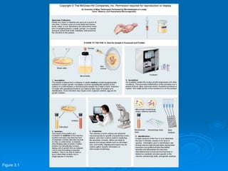
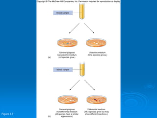
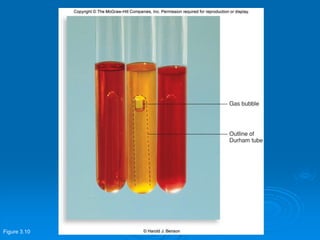
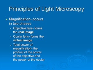
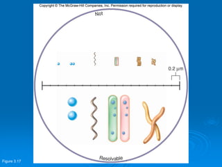
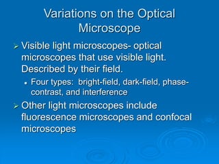
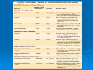
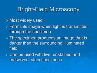
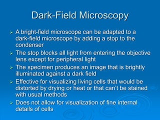
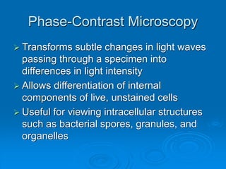
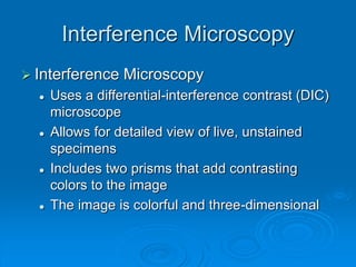
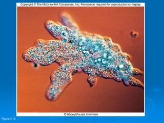
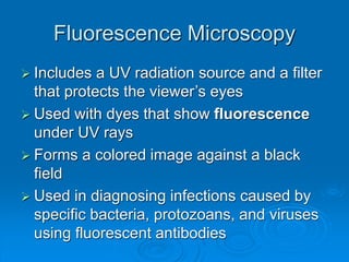
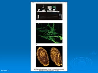
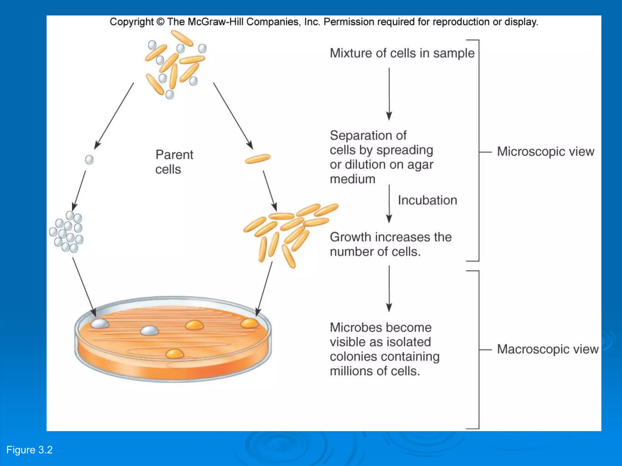
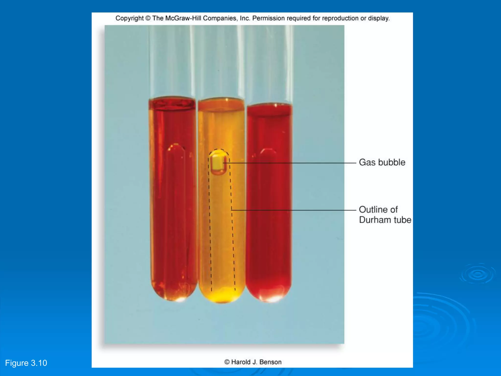
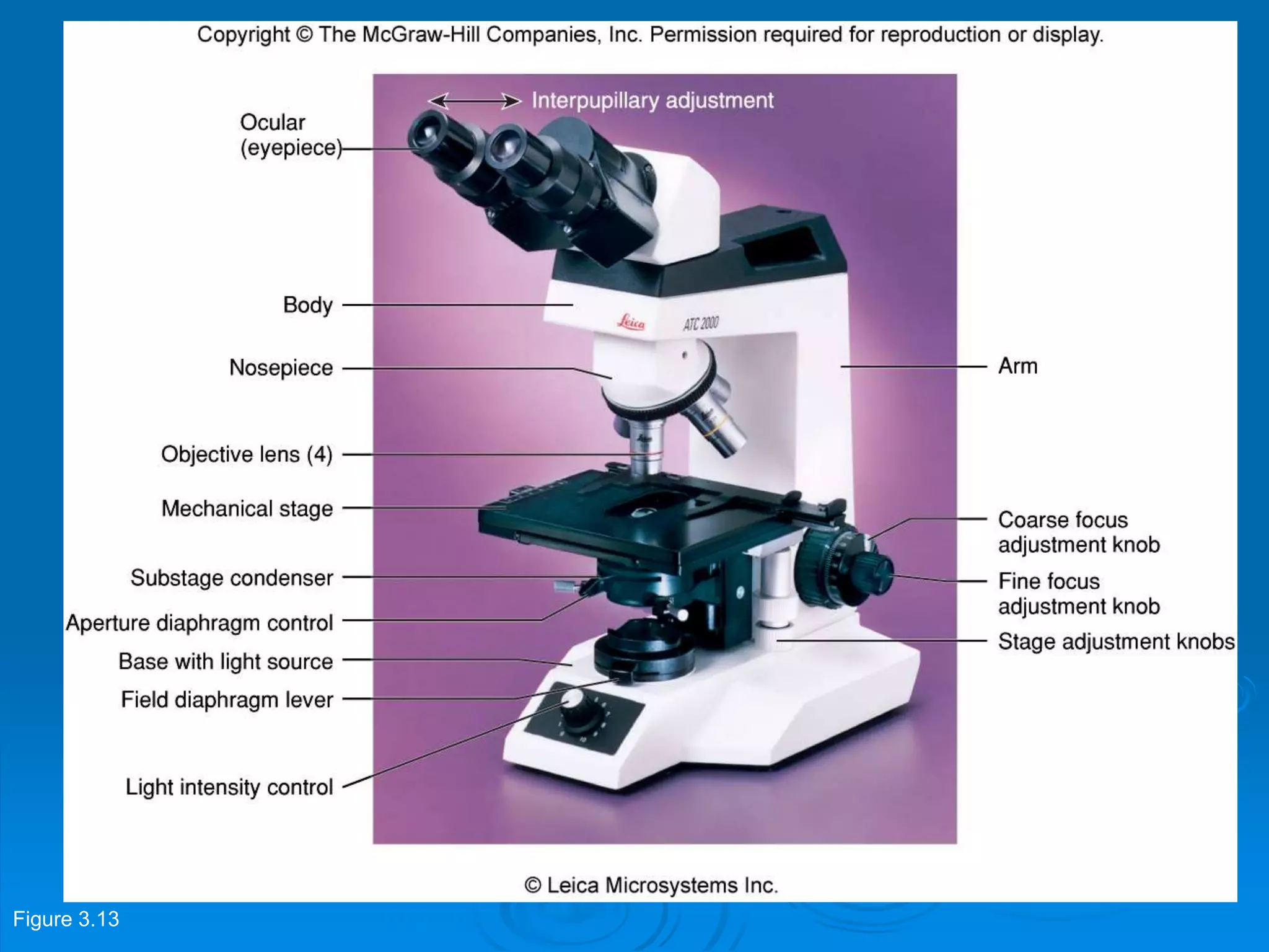
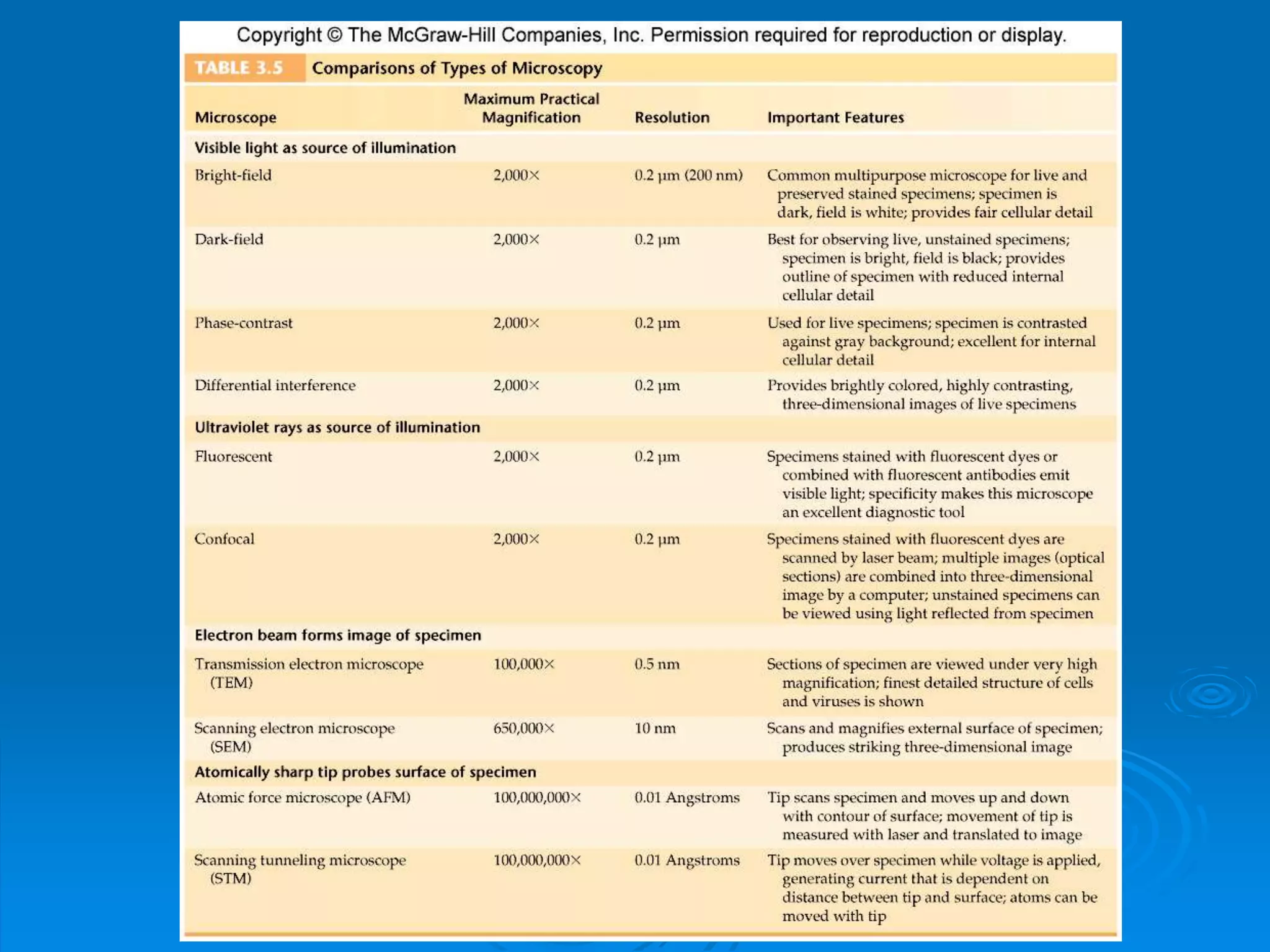
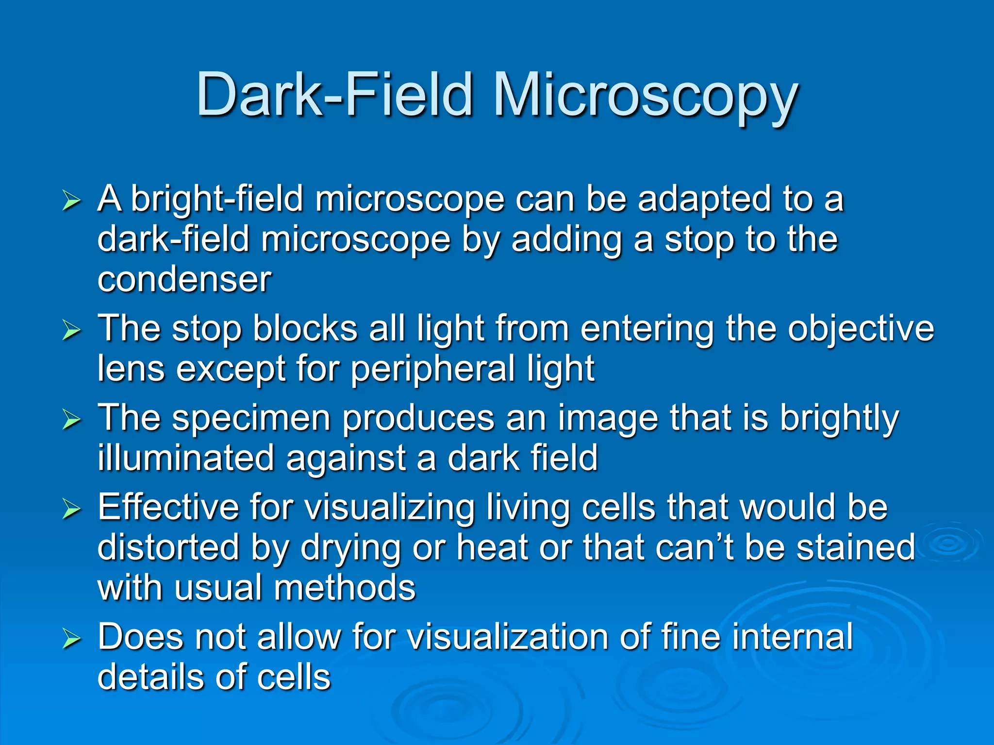
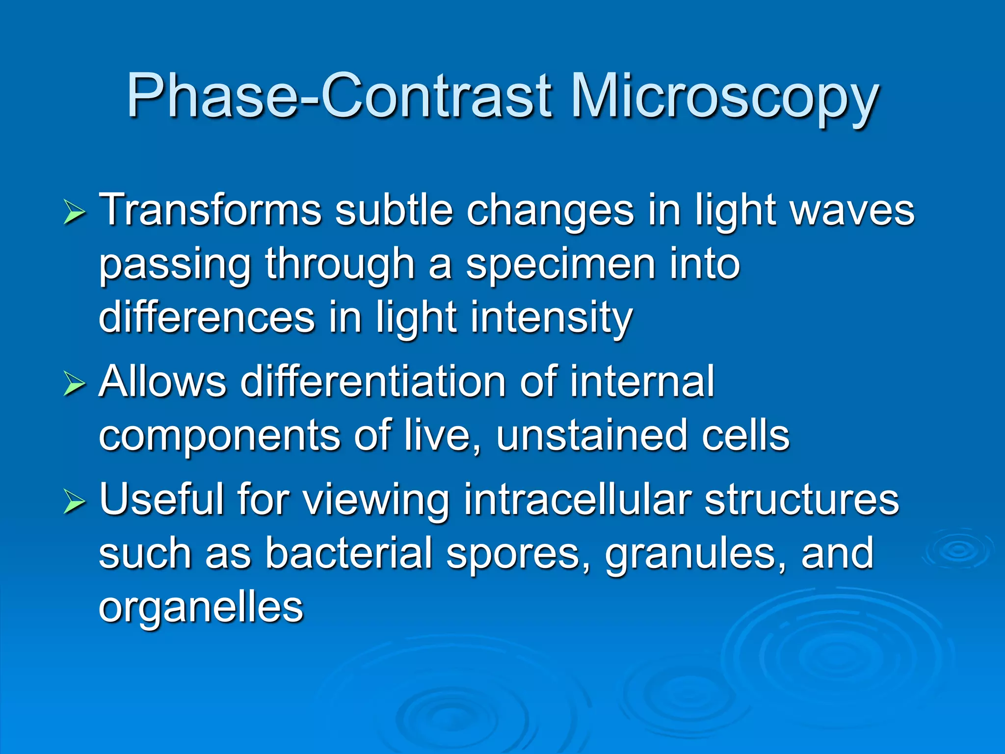
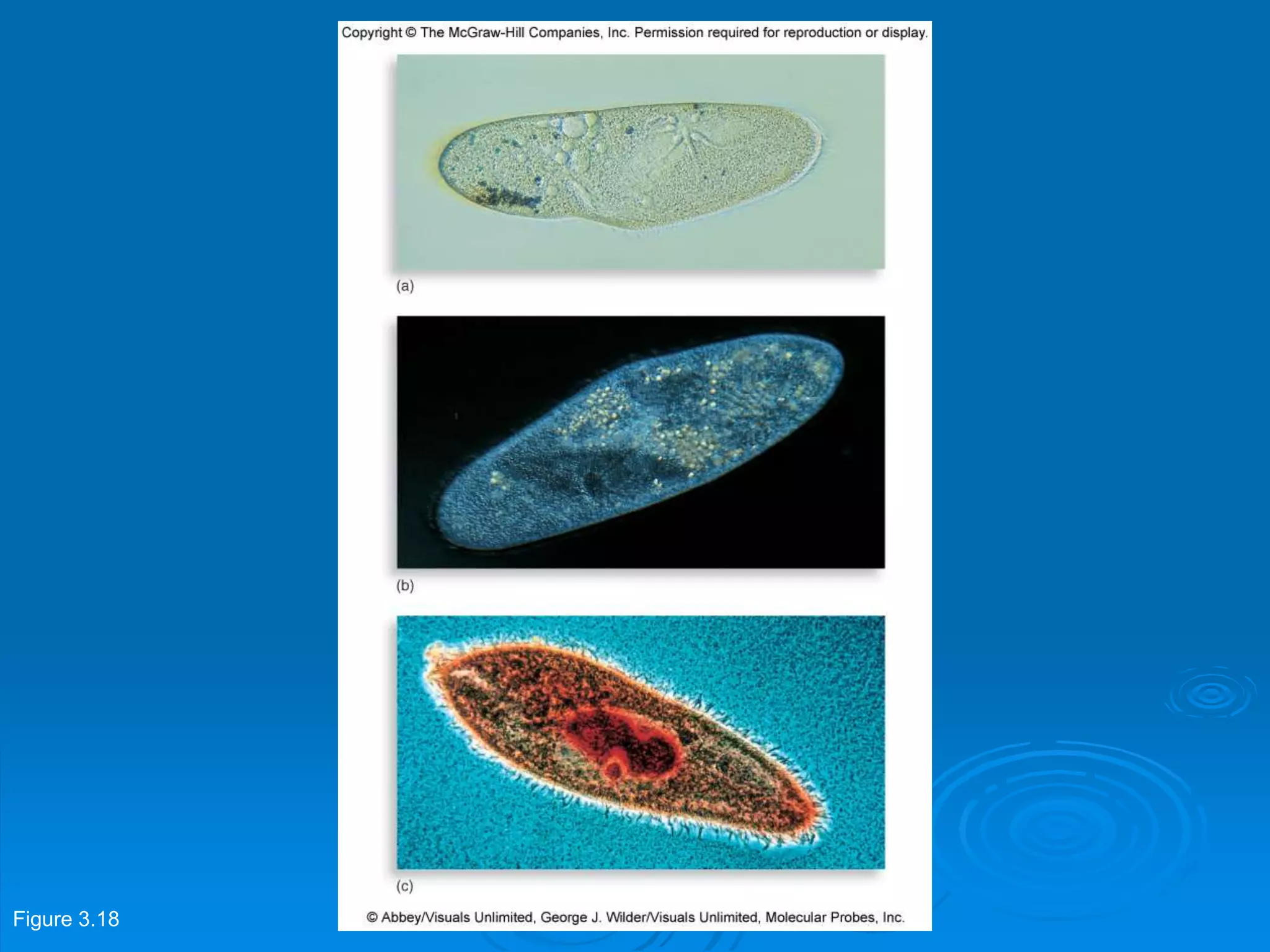
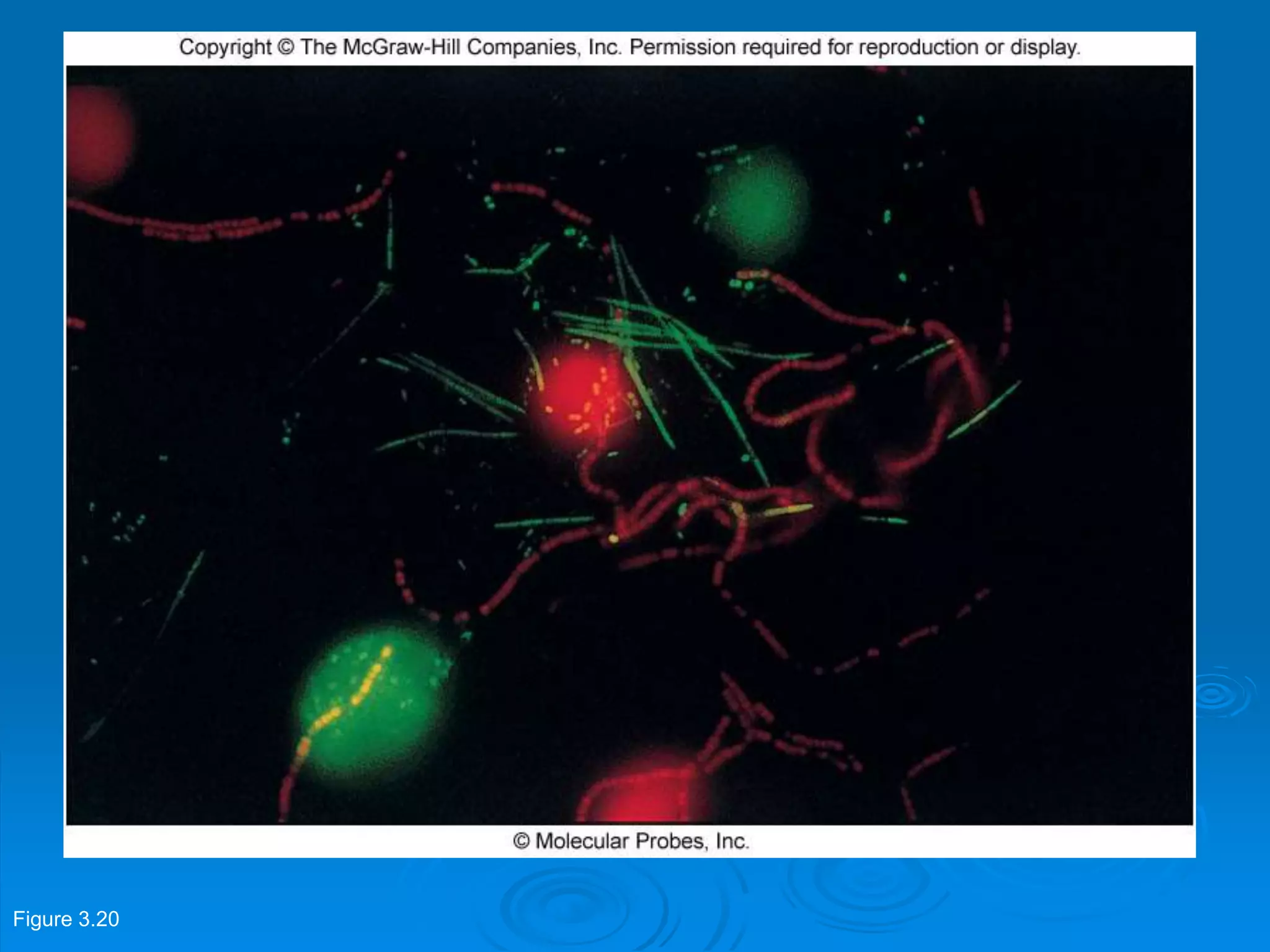
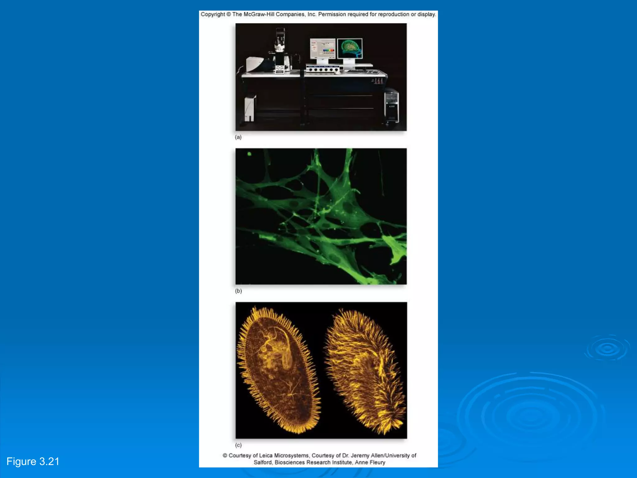
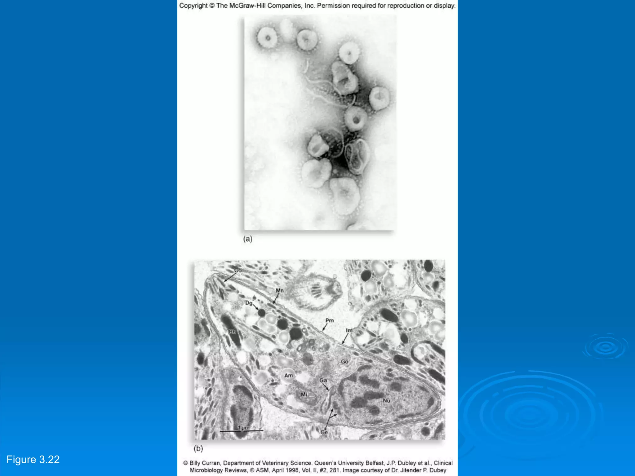
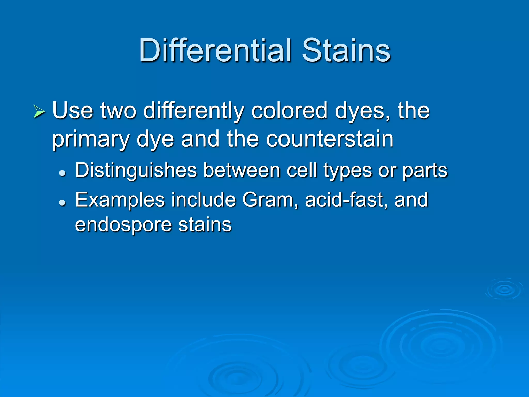
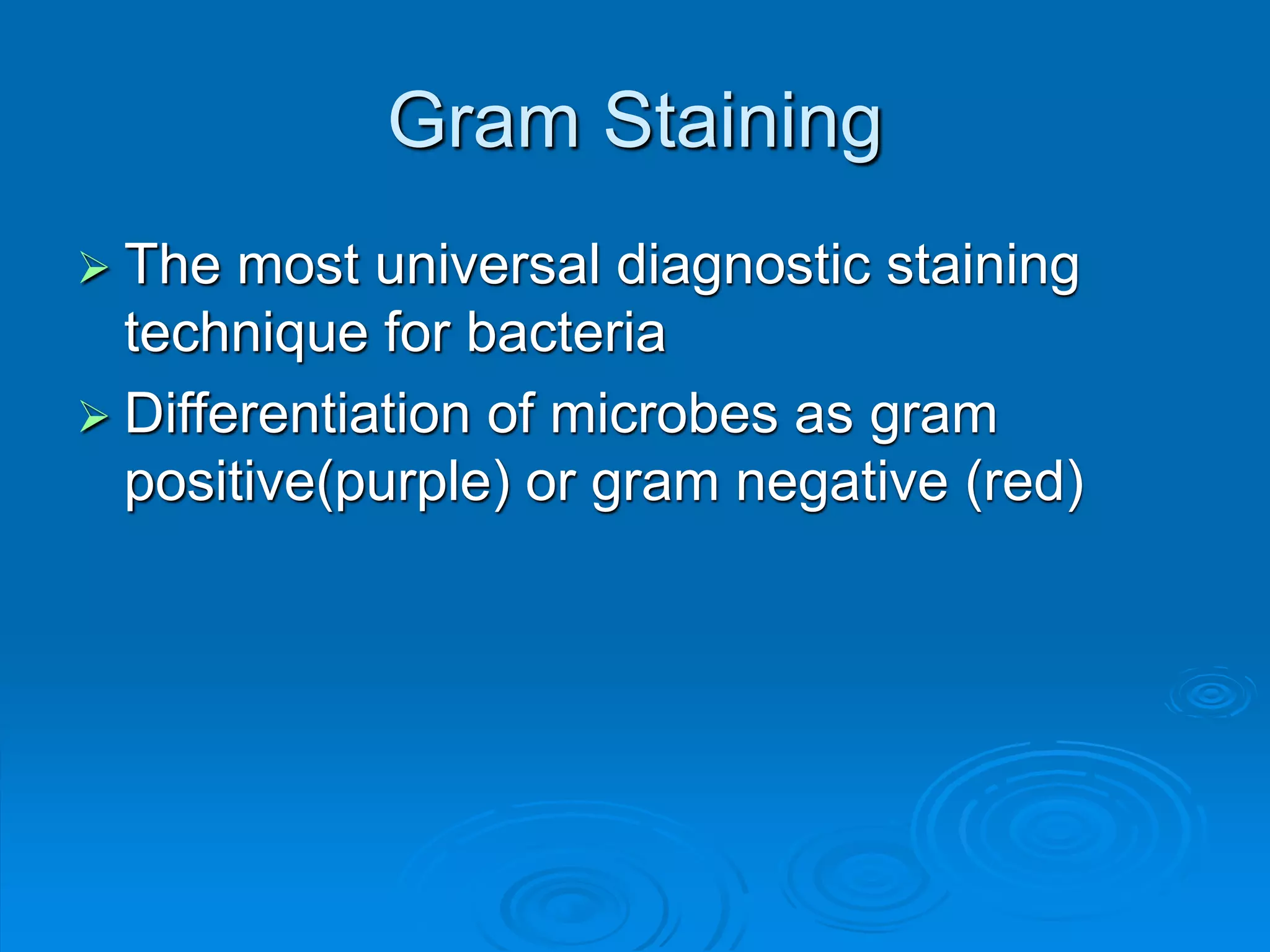
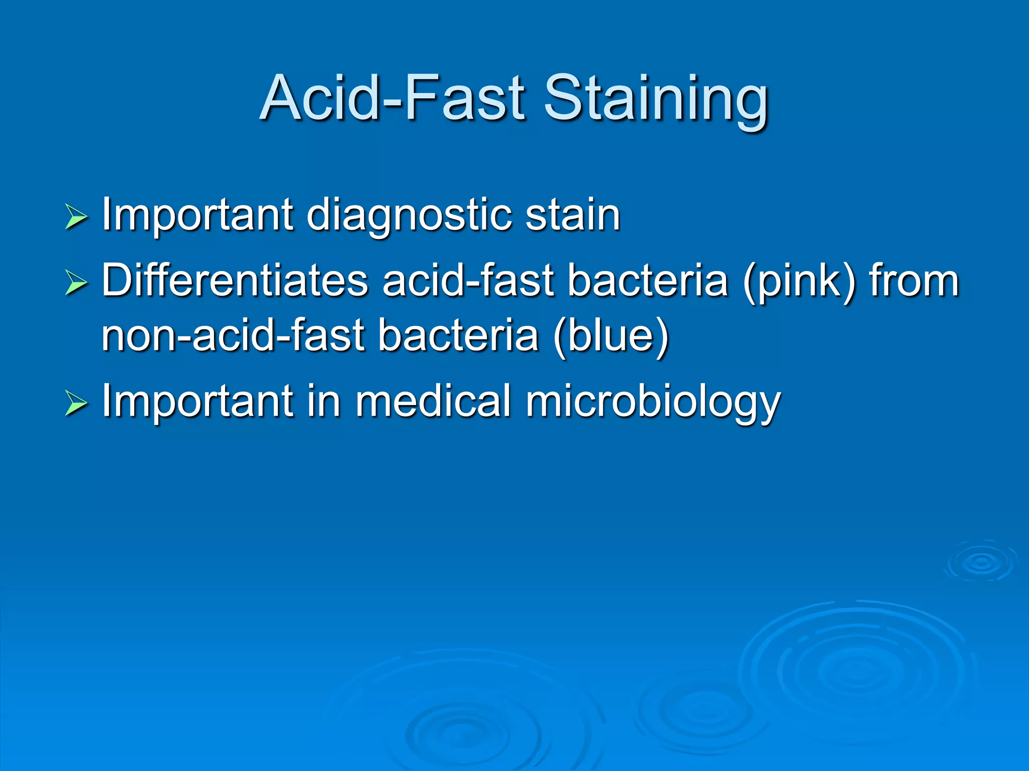
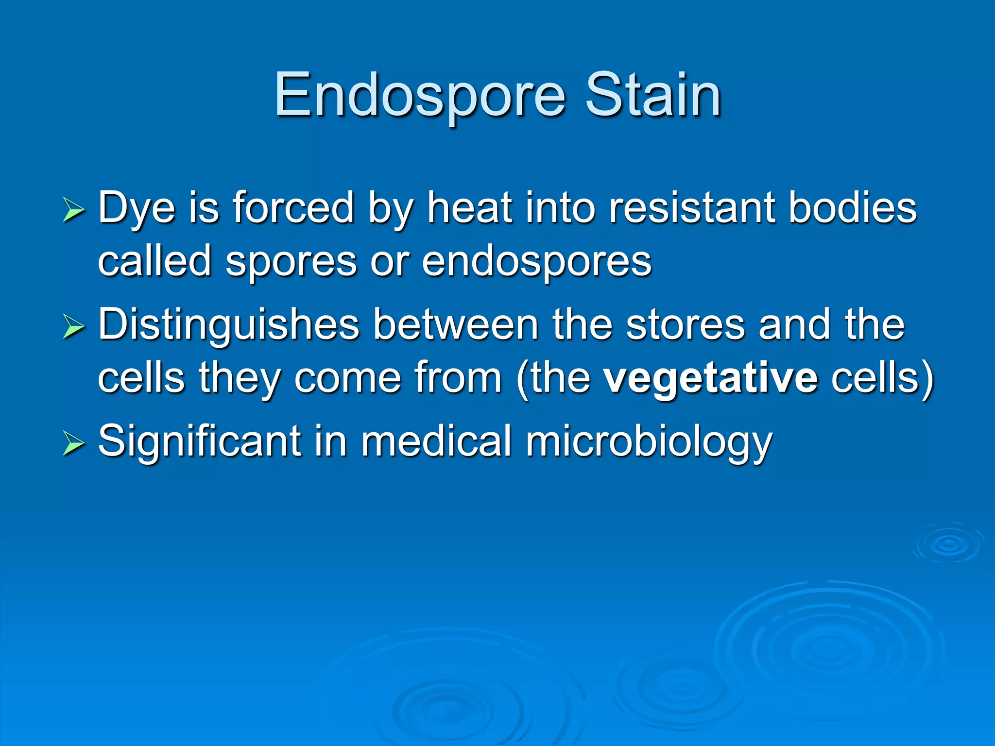
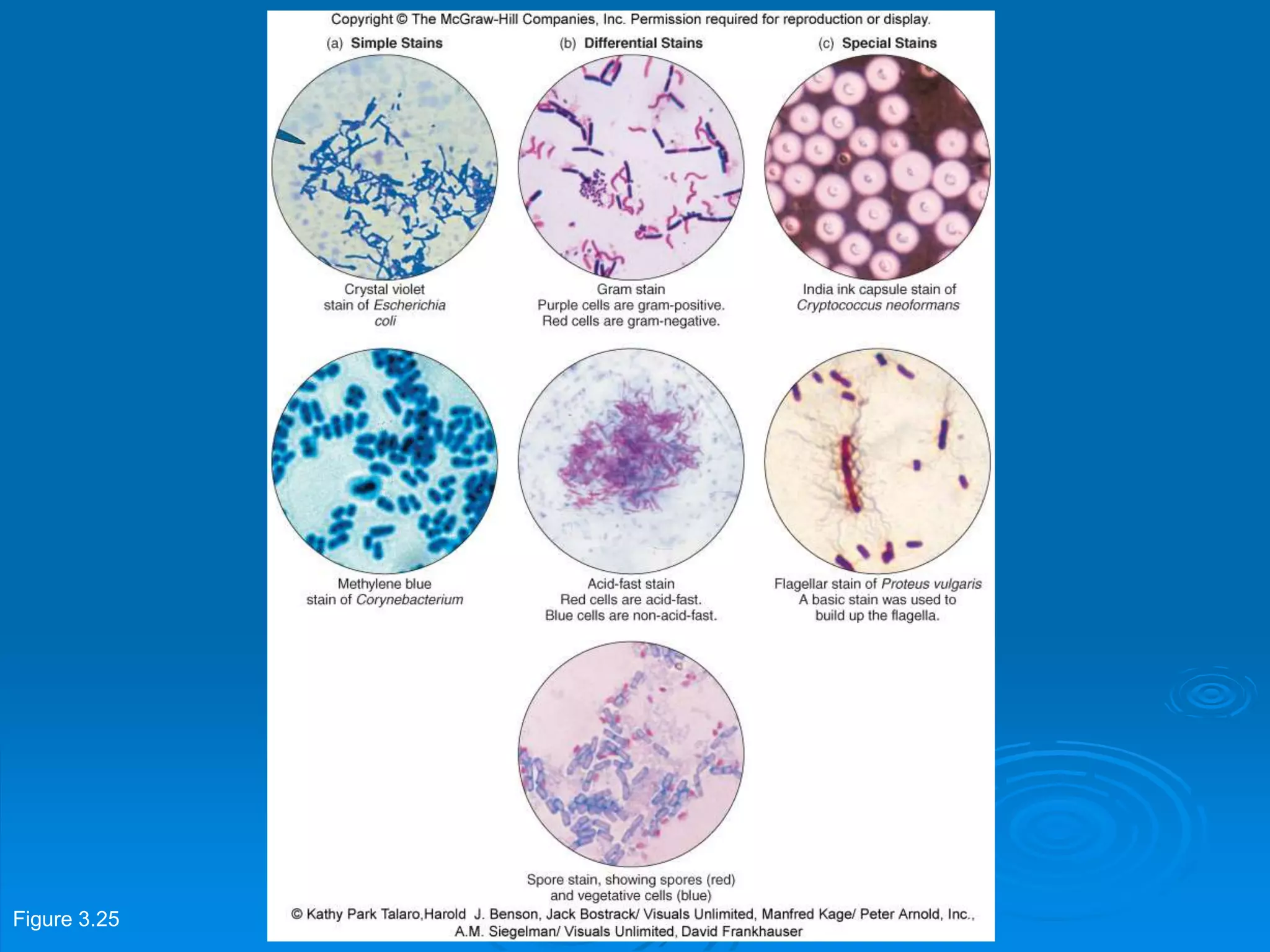
KEMBAR78
chapt03_lecture.ppt
Microbiology: A
Systems Approach, 2nd
ed.
Chapter 3: Tools of the
Laboratory
3.1 Methods of Culturing
Microorganisms: The Five I’s
 Microbiologists use five basic techniques
to manipulate, grow, examine, and
characterize microorganisms in the
laboratory: inoculation, incubation,
isolation, inspection, and identification
Figure 3.1
Inoculation and Isolation
 Inoculation: producing a culture
 Introduce a tiny sample (the inoculums) into a
container of nutrient medium
 Isolation: separating one species from
another
 Separating a single bacterial cell from other cells
and providing it space on a nutrient surface will
allow that cell to grow in to a mound of cells (a
colony).
 If formed from a single cell, the colony contains
cells from just that species.
Figure 3.2
Streak Plate Method
 Streak plate method- small droplet of culture
or sample spread over surface of the medium
with an inoculating loop
 Uses a pattern that thins out the sample and
separates the cells
Figure 3.3 a,b
Loop Dilation Method
 Loop dilation, or pour plate, method- sample
inoculated serially in to a series of liquid agar tues
to dilute the number of cells in each successive
tubes
 Tubes are then poured in to sterile Petri dishes and
allowed to solidify
Figure 3.3 c,d
Spread Plate Method
• Spread plate method- small volume of liquid, diluted
sample pipette on to surface of the medium and spread
around evenly by a sterile spreading tool
Figure 3.3 e,f
Media: Providing Nutrients in the
Laboratory
 At least 500 different types
 Contained in test tubes, flasks, or Petri
dishes
 Inoculated by loops, needles, pipettes, and
swabs
 Sterile technique necessary
 Classification of media
 Physical state
 Chemical composition
 Functional type
Classification of Media by Physical
State
 Liquid media: water-based solutions, do not solidify at
temperatures above freezing, flow freely when
container is tilted
 Broths, milks, or infusions
 Growth seen as cloudiness or particulates
 Semisolid media: clotlike consistency at room
temperature
 Used to determine motility and to localize reactions at a
specific site
 Solid media: a firm surface on which cells can form
discrete colonies
 Liquefiable and nonliquefiable
 Useful for isolating and culturing bacteria and fungi
Figure 3.4
Classification of Media by Chemical
Content
 Synthetic media- compositions are
precisely chemically defined
 Complex (nonsynthetic) media- if even just
one component is not chemically definable
Classification of Media by
Function
 General purpose media- to grow as broad
a spectrum of microbes as possible
 Usually nonsynthetic
 Contain a mixture of nutrients to support a
variety of microbes
 Examples: nutrient agar and broth, brain-
heart infusion, trypticase soy agar (TSA).
Enriched Media
• Enriched media- contain complex organic
substances (for example blood, serum,
growth factors) to support the growth of
fastidious bacteria. Examples: blood
agar, Thayer-Martin medium (chocolate
agar)
Figure 3.6
Selective and Differential Media
 Selective media- contains one or more
agents that inhibit the growth of certain
microbes but not others. Example: Mannitol
salt agar (MSA), MacConkey agar, Hektoen
enteric (HE) agar.
 Differential media- allow multiple types of
microorganisms to grow but display visible
differences among those microorganisms.
MacConkey agar can be used as a
differential medium as well.
Figure 3.7
Figure 3.8
Figure 3.9
Miscellaneous Media
 Reducing media- absorbs oxygen or slows its
penetration in the medium; used for growing
anaerobes or for determining oxygen requirements
 Carbohydrate fermentation media- contain sugars
that can be fermented and a pH indicator; useful
for identification of microorganisms
 Transport media- used to maintain and preserve
specimens that need to be held for a period of
time
 Assay media- used to test the effectiveness of
antibiotics, disinfectants, antiseptics, etc.
 Enumeration media- used to count the numbers of
organisms in a sample.
Figure 3.10
Incubation
 Incubation: an inoculated sample is placed in an
incubator to encourage growth.
 Usually in laboratories, between 20° and 40°C.
 Can control atmospheric gases as well.
 Can visually recognize growth as cloudiness in liquid
media and colonies on solid media.
 Pure culture- growth of only a single known species
(also called axenic)
• Usually created by subculture
 Mixed culture- holds two or more identified species
 Contaminated culture- includes unwanted
microorganisms of uncertain identity, or
contaminants.
Inspection and Identification
• Inspection and identification: Using appearance
as well as metabolism (biochemical tests) and
sometimes genetic analysis or immunologic
testing to identify the organisms in a culture.
 Cultures can be maintained using stock
cultures
 Once cultures are no longer being used,
they must be sterilized and destroyed
properly.
3.2 The Microscope: Window on
an Invisible Realm
 Two key characteristics of microscopes:
magnification and resolving power
 Magnification
 Results when visible light waves pass through a
curved lens
 The light experiences refraction
 An image is formed by the refracted light when an
object is placed a certain distance from the lens
and is illuminated with light
 The image is enlarged to a particular degree- the
power of magnification
Figure 3.13
Principles of Light Microscopy
 Magnification- occurs
in two phases
 Objective lens- forms
the real image
 Ocular lens- forms the
virtual image
 Total power of
magnification- the
product of the power
of the objective and
the power of the ocular
Resolution
 Resolution- the ability to distinguish two adjacent
objects or points from one another
 Also known as resolving power
 Resolving power (RP) = Wavelength of light in nm
2 x Numerical aperture of
objective lens
 Resolution distance= 0.61 x wavelength of light in nm
Numerical aperture of objective
lens
 Shorter wavelengths provide a better resolution
 Numerical aperture- describes the relative efficiency
Figure 3.15
Figure 3.16
Magnification and Resolution
 Increased magnification decreases the
resolution
• Adjusting the amount of light entering the
condenser using an adjustable iris
diaphragm or using special dyes help
increase resolution at higher
magnifications
Figure 3.17
Variations on the Optical
Microscope
 Visible light microscopes- optical
microscopes that use visible light.
Described by their field.
 Four types: bright-field, dark-field, phase-
contrast, and interference
 Other light microscopes include
fluorescence microscopes and confocal
microscopes
Bright-Field Microscopy
 Most widely used
 Forms its image when light is transmitted
through the specimen
 The specimen produces an image that is
darker than the surrounding illuminated
field
 Can be used with live, unstained and
preserved, stain specimens
Dark-Field Microscopy
 A bright-field microscope can be adapted to a
dark-field microscope by adding a stop to the
condenser
 The stop blocks all light from entering the objective
lens except for peripheral light
 The specimen produces an image that is brightly
illuminated against a dark field
 Effective for visualizing living cells that would be
distorted by drying or heat or that can’t be stained
with usual methods
 Does not allow for visualization of fine internal
details of cells
Phase-Contrast Microscopy
 Transforms subtle changes in light waves
passing through a specimen into
differences in light intensity
 Allows differentiation of internal
components of live, unstained cells
 Useful for viewing intracellular structures
such as bacterial spores, granules, and
organelles
Figure 3.18
Interference Microscopy
 Interference Microscopy
 Uses a differential-interference contrast (DIC)
microscope
 Allows for detailed view of live, unstained
specimens
 Includes two prisms that add contrasting
colors to the image
 The image is colorful and three-dimensional
Figure 3.19
Fluorescence Microscopy
 Includes a UV radiation source and a filter
that protects the viewer’s eyes
 Used with dyes that show fluorescence
under UV rays
 Forms a colored image against a black
field
 Used in diagnosing infections caused by
specific bacteria, protozoans, and viruses
using fluorescent antibodies
Figure 3.20
Confocal Microscopy
 Allows for viewing cells at higher
magnifications using a laser beam of light
to scan various depths in the specimen
 Most often used on fluorescently stained
specimens
Figure 3.21
Electron Microscopy
 Originally developed for studying nonbiological
materials
 Biologists began using it in the early 1930s
 Forms an image with a beam of electrons
 Electrons travel in wavelike patterns 1,000 times
shorter than visible light waves
 This increases the resolving power tremendously
 Magnification can be extremely high (between
5,000X and 1,000,000X for biological specimens)
 Allows scientists to view the finest structure of
cells
 Two forms: transmission electron microscope
(TEM) and scanning electron microscope (SEM)
TEM
 Often used to view structures of cells and
viruses
 Electrons are transmitted through the
specimen
 The specimen must be very thin (20-100
nm thick) and stained to increase image
contrast
 Dark areas of a TEM image represent
thicker or denser parts
Figure 3.22
SEM
 Creates an extremely detailed three-
dimensional view of all kinds of objects
 Electrons bombard the surface of a whole
metal-coated specimen
 Electrons deflected from the surface are
picked up by a sophisticated detector
 The electron pattern is displayed as an
image on a television screen
 Contours of specimens resolved with SEM
are very revealing and surprising
Figure 3.23
Preparing Specimens for Optical
Microscopes
 Generally prepared by mounting a sample
on a glass slide
 How the slide is prepared depends on
 The condition of the specimen (living or
preserved)
 The aims of the examiner (to observe overall
structure, identify microorganisms, or see
movement)
 The type of microscopy available
Living Preparations
 Wet mounts or hanging drop mounts
 Wet mount:
 Cells suspended in fluid, a drop or two of the
culture is then placed on a slide and overlaid with
a cover glass
 Cover glass can damage larger cells and might
dry or contaminate the observer’s fingers
 Hanging drop mount:
 Uses a depression slide, Vaseline, and coverslip
 The sample is suspended from the coverslip
Figure 3.24
Fixed, Stained Smears
 Smear technique developed by Robert Koch
 Spread a thin film made from a liquid suspension of
cells and air-drying it
 Heat the dried smear by a process called heat fixation
 Some cells are fixed using chemicals
 Staining creates contrast and allows features of
the cells to stand out
 Applies colored chemicals to specimens
 Dyes become affixed to the cells through a chemical
reaction
 Dyes are classified as basic (cationic) dyes, or acidic
(anionic) dyes.
Positive and Negative Staining
 Positive staining: the dye sticks to the
specimen to give it color
 Negative staining: The dye does not stick to
the specimen, instead settles around its
boundaries, creating a silhouette.
 Nigrosin and India ink commonly used
 Heat fixation not required, so there is less
shrinkage or distortion of cells
 Also used to accentuate the capsule surrounding
certain bacteria and yeasts
Simple Stains
 Require only a single dye
 Examples include malachite green, crystal
violet, basic fuchsin, and safranin
 All cells appear the same color but can reveal
shape, size, and arrangement
Differential Stains
 Use two differently colored dyes, the
primary dye and the counterstain
 Distinguishes between cell types or parts
 Examples include Gram, acid-fast, and
endospore stains
Gram Staining
 The most universal diagnostic staining
technique for bacteria
 Differentiation of microbes as gram
positive(purple) or gram negative (red)
Acid-Fast Staining
 Important diagnostic stain
 Differentiates acid-fast bacteria (pink) from
non-acid-fast bacteria (blue)
 Important in medical microbiology
Endospore Stain
 Dye is forced by heat into resistant bodies
called spores or endospores
 Distinguishes between the stores and the
cells they come from (the vegetative cells)
 Significant in medical microbiology
Special Stains
 Used to emphasize certain cell parts that
aren’t revealed by conventional staining
methods
 Examples: capsule staining, flagellar
staining
Figure 3.25

chapt03_lecture.ppt

  • 1.
    Microbiology: A Systems Approach,2nd ed. Chapter 3: Tools of the Laboratory
  • 2.
    3.1 Methods ofCulturing Microorganisms: The Five I’s  Microbiologists use five basic techniques to manipulate, grow, examine, and characterize microorganisms in the laboratory: inoculation, incubation, isolation, inspection, and identification
  • 3.
  • 4.
    Inoculation and Isolation Inoculation: producing a culture  Introduce a tiny sample (the inoculums) into a container of nutrient medium  Isolation: separating one species from another  Separating a single bacterial cell from other cells and providing it space on a nutrient surface will allow that cell to grow in to a mound of cells (a colony).  If formed from a single cell, the colony contains cells from just that species.
  • 5.
  • 6.
    Streak Plate Method Streak plate method- small droplet of culture or sample spread over surface of the medium with an inoculating loop  Uses a pattern that thins out the sample and separates the cells Figure 3.3 a,b
  • 7.
    Loop Dilation Method Loop dilation, or pour plate, method- sample inoculated serially in to a series of liquid agar tues to dilute the number of cells in each successive tubes  Tubes are then poured in to sterile Petri dishes and allowed to solidify Figure 3.3 c,d
  • 8.
    Spread Plate Method •Spread plate method- small volume of liquid, diluted sample pipette on to surface of the medium and spread around evenly by a sterile spreading tool Figure 3.3 e,f
  • 9.
    Media: Providing Nutrientsin the Laboratory  At least 500 different types  Contained in test tubes, flasks, or Petri dishes  Inoculated by loops, needles, pipettes, and swabs  Sterile technique necessary  Classification of media  Physical state  Chemical composition  Functional type
  • 11.
    Classification of Mediaby Physical State  Liquid media: water-based solutions, do not solidify at temperatures above freezing, flow freely when container is tilted  Broths, milks, or infusions  Growth seen as cloudiness or particulates  Semisolid media: clotlike consistency at room temperature  Used to determine motility and to localize reactions at a specific site  Solid media: a firm surface on which cells can form discrete colonies  Liquefiable and nonliquefiable  Useful for isolating and culturing bacteria and fungi
  • 12.
  • 13.
    Classification of Mediaby Chemical Content  Synthetic media- compositions are precisely chemically defined  Complex (nonsynthetic) media- if even just one component is not chemically definable
  • 14.
    Classification of Mediaby Function  General purpose media- to grow as broad a spectrum of microbes as possible  Usually nonsynthetic  Contain a mixture of nutrients to support a variety of microbes  Examples: nutrient agar and broth, brain- heart infusion, trypticase soy agar (TSA).
  • 15.
    Enriched Media • Enrichedmedia- contain complex organic substances (for example blood, serum, growth factors) to support the growth of fastidious bacteria. Examples: blood agar, Thayer-Martin medium (chocolate agar)
  • 16.
  • 17.
    Selective and DifferentialMedia  Selective media- contains one or more agents that inhibit the growth of certain microbes but not others. Example: Mannitol salt agar (MSA), MacConkey agar, Hektoen enteric (HE) agar.  Differential media- allow multiple types of microorganisms to grow but display visible differences among those microorganisms. MacConkey agar can be used as a differential medium as well.
  • 18.
  • 19.
  • 20.
  • 21.
    Miscellaneous Media  Reducingmedia- absorbs oxygen or slows its penetration in the medium; used for growing anaerobes or for determining oxygen requirements  Carbohydrate fermentation media- contain sugars that can be fermented and a pH indicator; useful for identification of microorganisms  Transport media- used to maintain and preserve specimens that need to be held for a period of time  Assay media- used to test the effectiveness of antibiotics, disinfectants, antiseptics, etc.  Enumeration media- used to count the numbers of organisms in a sample.
  • 22.
  • 23.
    Incubation  Incubation: aninoculated sample is placed in an incubator to encourage growth.  Usually in laboratories, between 20° and 40°C.  Can control atmospheric gases as well.  Can visually recognize growth as cloudiness in liquid media and colonies on solid media.  Pure culture- growth of only a single known species (also called axenic) • Usually created by subculture  Mixed culture- holds two or more identified species  Contaminated culture- includes unwanted microorganisms of uncertain identity, or contaminants.
  • 24.
    Inspection and Identification •Inspection and identification: Using appearance as well as metabolism (biochemical tests) and sometimes genetic analysis or immunologic testing to identify the organisms in a culture.  Cultures can be maintained using stock cultures  Once cultures are no longer being used, they must be sterilized and destroyed properly.
  • 25.
    3.2 The Microscope:Window on an Invisible Realm  Two key characteristics of microscopes: magnification and resolving power  Magnification  Results when visible light waves pass through a curved lens  The light experiences refraction  An image is formed by the refracted light when an object is placed a certain distance from the lens and is illuminated with light  The image is enlarged to a particular degree- the power of magnification
  • 26.
  • 27.
    Principles of LightMicroscopy  Magnification- occurs in two phases  Objective lens- forms the real image  Ocular lens- forms the virtual image  Total power of magnification- the product of the power of the objective and the power of the ocular
  • 28.
    Resolution  Resolution- theability to distinguish two adjacent objects or points from one another  Also known as resolving power  Resolving power (RP) = Wavelength of light in nm 2 x Numerical aperture of objective lens  Resolution distance= 0.61 x wavelength of light in nm Numerical aperture of objective lens  Shorter wavelengths provide a better resolution  Numerical aperture- describes the relative efficiency
  • 29.
  • 30.
  • 31.
    Magnification and Resolution Increased magnification decreases the resolution • Adjusting the amount of light entering the condenser using an adjustable iris diaphragm or using special dyes help increase resolution at higher magnifications
  • 32.
  • 33.
    Variations on theOptical Microscope  Visible light microscopes- optical microscopes that use visible light. Described by their field.  Four types: bright-field, dark-field, phase- contrast, and interference  Other light microscopes include fluorescence microscopes and confocal microscopes
  • 35.
    Bright-Field Microscopy  Mostwidely used  Forms its image when light is transmitted through the specimen  The specimen produces an image that is darker than the surrounding illuminated field  Can be used with live, unstained and preserved, stain specimens
  • 36.
    Dark-Field Microscopy  Abright-field microscope can be adapted to a dark-field microscope by adding a stop to the condenser  The stop blocks all light from entering the objective lens except for peripheral light  The specimen produces an image that is brightly illuminated against a dark field  Effective for visualizing living cells that would be distorted by drying or heat or that can’t be stained with usual methods  Does not allow for visualization of fine internal details of cells
  • 37.
    Phase-Contrast Microscopy  Transformssubtle changes in light waves passing through a specimen into differences in light intensity  Allows differentiation of internal components of live, unstained cells  Useful for viewing intracellular structures such as bacterial spores, granules, and organelles
  • 38.
  • 39.
    Interference Microscopy  InterferenceMicroscopy  Uses a differential-interference contrast (DIC) microscope  Allows for detailed view of live, unstained specimens  Includes two prisms that add contrasting colors to the image  The image is colorful and three-dimensional
  • 40.
  • 41.
    Fluorescence Microscopy  Includesa UV radiation source and a filter that protects the viewer’s eyes  Used with dyes that show fluorescence under UV rays  Forms a colored image against a black field  Used in diagnosing infections caused by specific bacteria, protozoans, and viruses using fluorescent antibodies
  • 42.
  • 43.
    Confocal Microscopy  Allowsfor viewing cells at higher magnifications using a laser beam of light to scan various depths in the specimen  Most often used on fluorescently stained specimens
  • 44.
  • 45.
    Electron Microscopy  Originallydeveloped for studying nonbiological materials  Biologists began using it in the early 1930s  Forms an image with a beam of electrons  Electrons travel in wavelike patterns 1,000 times shorter than visible light waves  This increases the resolving power tremendously  Magnification can be extremely high (between 5,000X and 1,000,000X for biological specimens)  Allows scientists to view the finest structure of cells  Two forms: transmission electron microscope (TEM) and scanning electron microscope (SEM)
  • 46.
    TEM  Often usedto view structures of cells and viruses  Electrons are transmitted through the specimen  The specimen must be very thin (20-100 nm thick) and stained to increase image contrast  Dark areas of a TEM image represent thicker or denser parts
  • 47.
  • 48.
    SEM  Creates anextremely detailed three- dimensional view of all kinds of objects  Electrons bombard the surface of a whole metal-coated specimen  Electrons deflected from the surface are picked up by a sophisticated detector  The electron pattern is displayed as an image on a television screen  Contours of specimens resolved with SEM are very revealing and surprising
  • 49.
  • 50.
    Preparing Specimens forOptical Microscopes  Generally prepared by mounting a sample on a glass slide  How the slide is prepared depends on  The condition of the specimen (living or preserved)  The aims of the examiner (to observe overall structure, identify microorganisms, or see movement)  The type of microscopy available
  • 51.
    Living Preparations  Wetmounts or hanging drop mounts  Wet mount:  Cells suspended in fluid, a drop or two of the culture is then placed on a slide and overlaid with a cover glass  Cover glass can damage larger cells and might dry or contaminate the observer’s fingers  Hanging drop mount:  Uses a depression slide, Vaseline, and coverslip  The sample is suspended from the coverslip
  • 52.
  • 53.
    Fixed, Stained Smears Smear technique developed by Robert Koch  Spread a thin film made from a liquid suspension of cells and air-drying it  Heat the dried smear by a process called heat fixation  Some cells are fixed using chemicals  Staining creates contrast and allows features of the cells to stand out  Applies colored chemicals to specimens  Dyes become affixed to the cells through a chemical reaction  Dyes are classified as basic (cationic) dyes, or acidic (anionic) dyes.
  • 55.
    Positive and NegativeStaining  Positive staining: the dye sticks to the specimen to give it color  Negative staining: The dye does not stick to the specimen, instead settles around its boundaries, creating a silhouette.  Nigrosin and India ink commonly used  Heat fixation not required, so there is less shrinkage or distortion of cells  Also used to accentuate the capsule surrounding certain bacteria and yeasts
  • 56.
    Simple Stains  Requireonly a single dye  Examples include malachite green, crystal violet, basic fuchsin, and safranin  All cells appear the same color but can reveal shape, size, and arrangement
  • 57.
    Differential Stains  Usetwo differently colored dyes, the primary dye and the counterstain  Distinguishes between cell types or parts  Examples include Gram, acid-fast, and endospore stains
  • 58.
    Gram Staining  Themost universal diagnostic staining technique for bacteria  Differentiation of microbes as gram positive(purple) or gram negative (red)
  • 59.
    Acid-Fast Staining  Importantdiagnostic stain  Differentiates acid-fast bacteria (pink) from non-acid-fast bacteria (blue)  Important in medical microbiology
  • 60.
    Endospore Stain  Dyeis forced by heat into resistant bodies called spores or endospores  Distinguishes between the stores and the cells they come from (the vegetative cells)  Significant in medical microbiology
  • 61.
    Special Stains  Usedto emphasize certain cell parts that aren’t revealed by conventional staining methods  Examples: capsule staining, flagellar staining
  • 62.