Downloaded 1,757 times

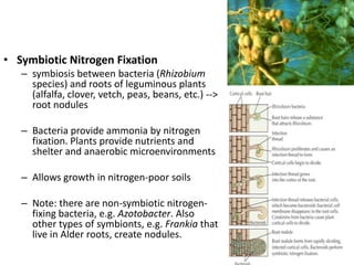
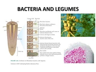


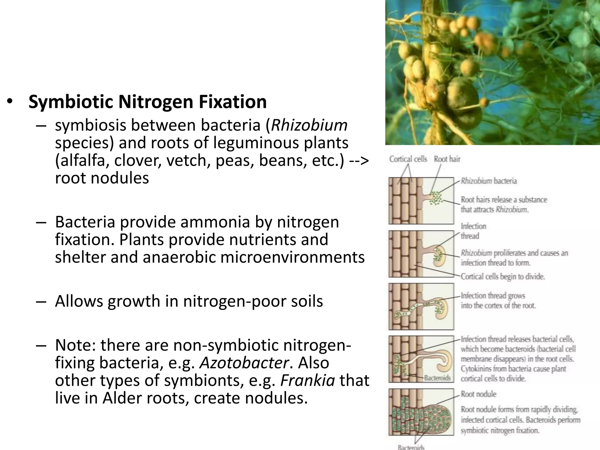


This document discusses microbial interactions, including both positive and negative interactions. It provides examples of symbiotic relationships between microbes and plants, such as lichens and nitrogen-fixing bacteria in legume roots. Microbial interactions with animals are also discussed, including relationships between rumen microbes and ruminant digestion. The document concludes by examining the interaction between the human microbiome and the host.
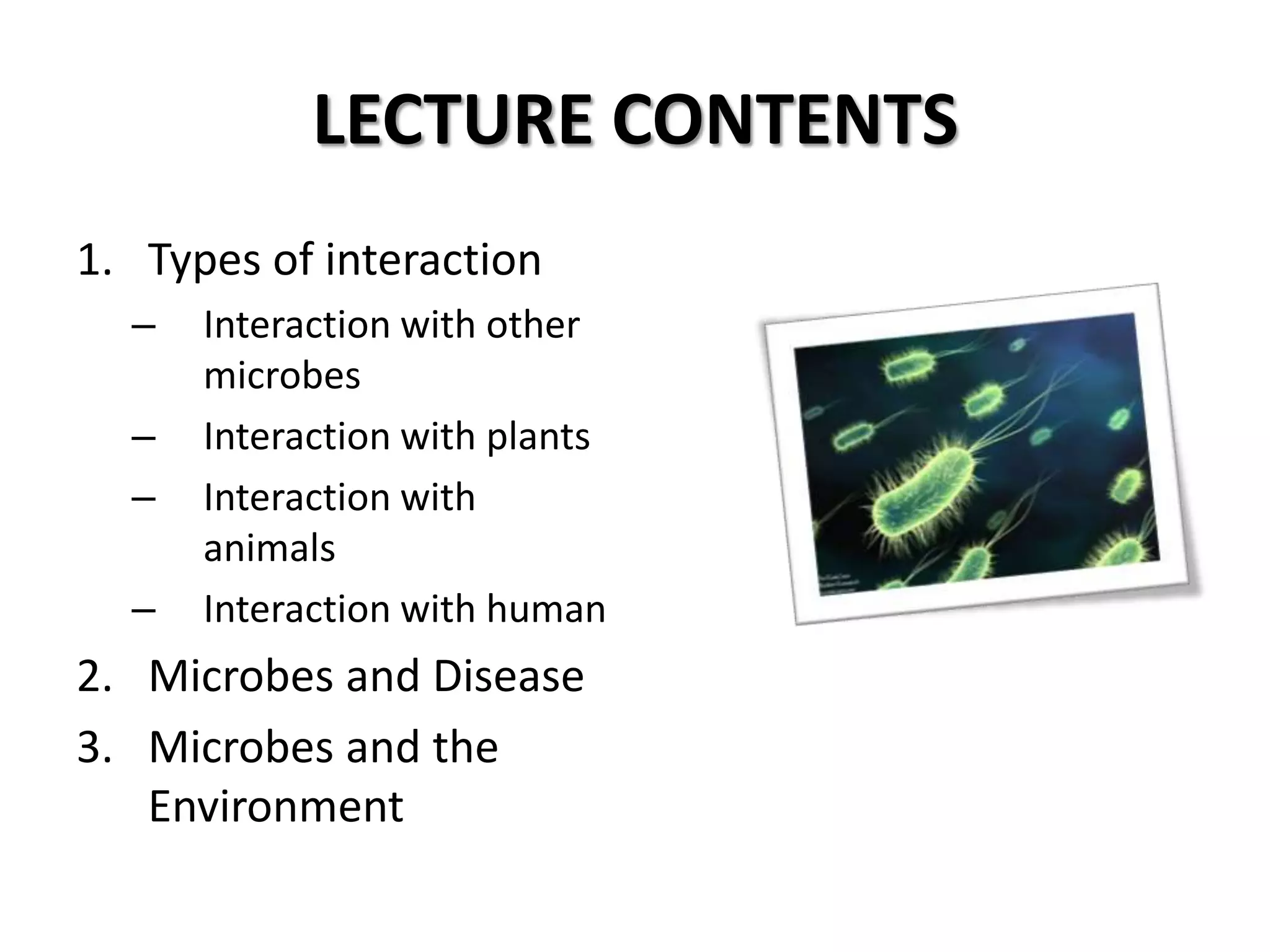
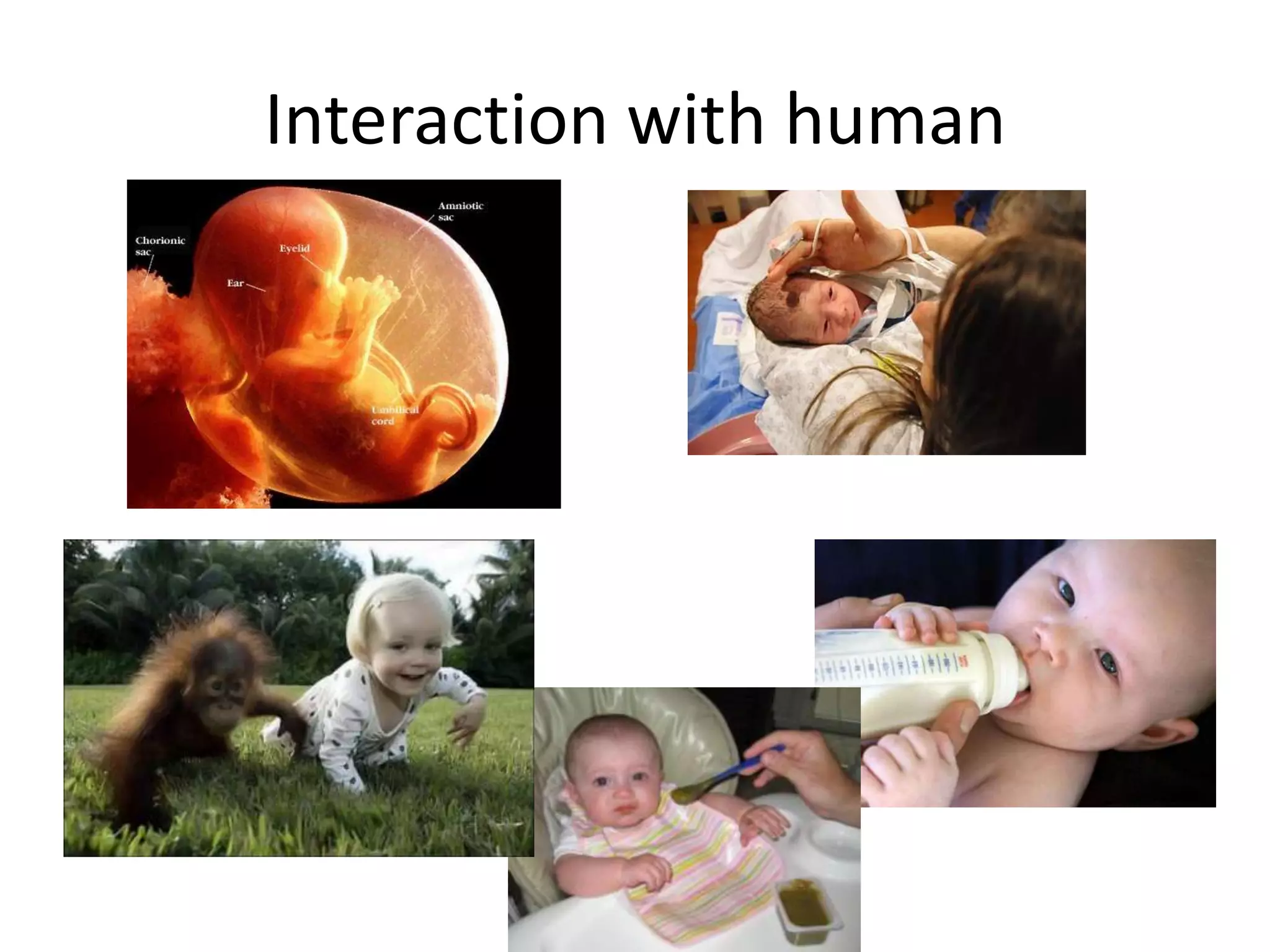
Overview of microbial interactions with other microbes, plants, animals, and humans, including pathogenic effects.
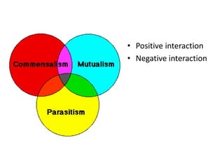
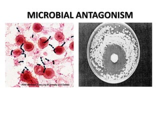
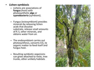
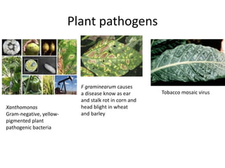
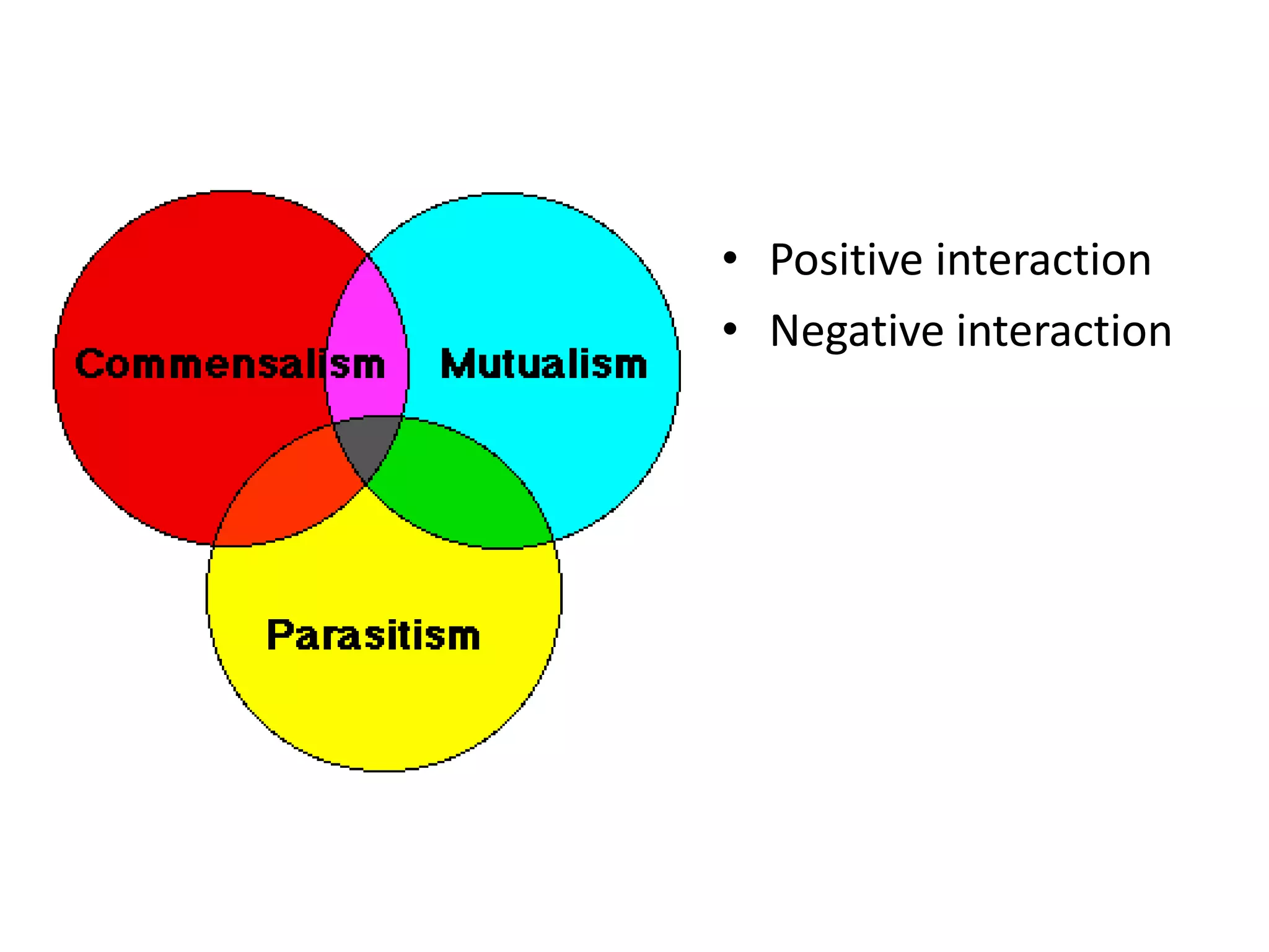
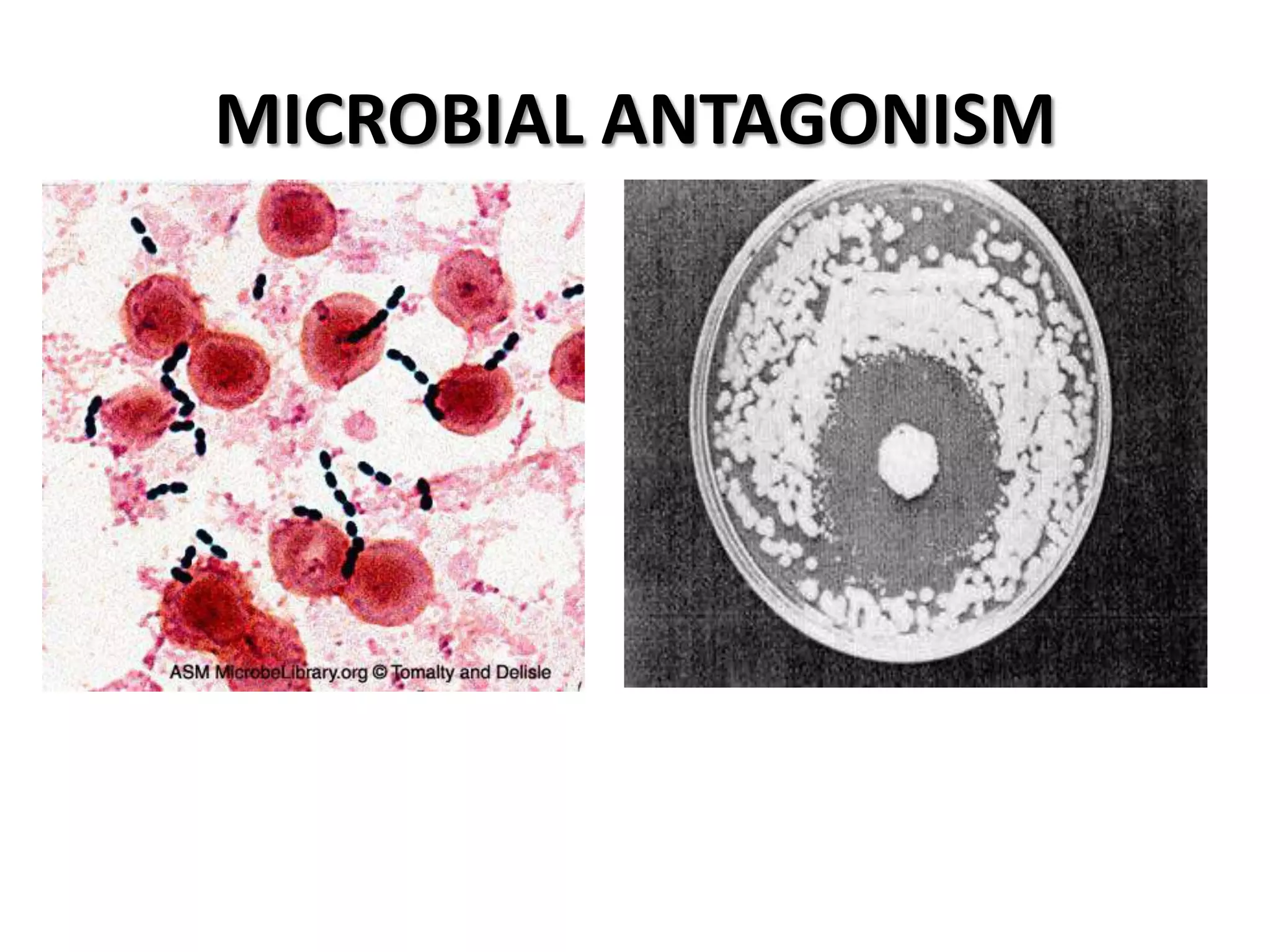
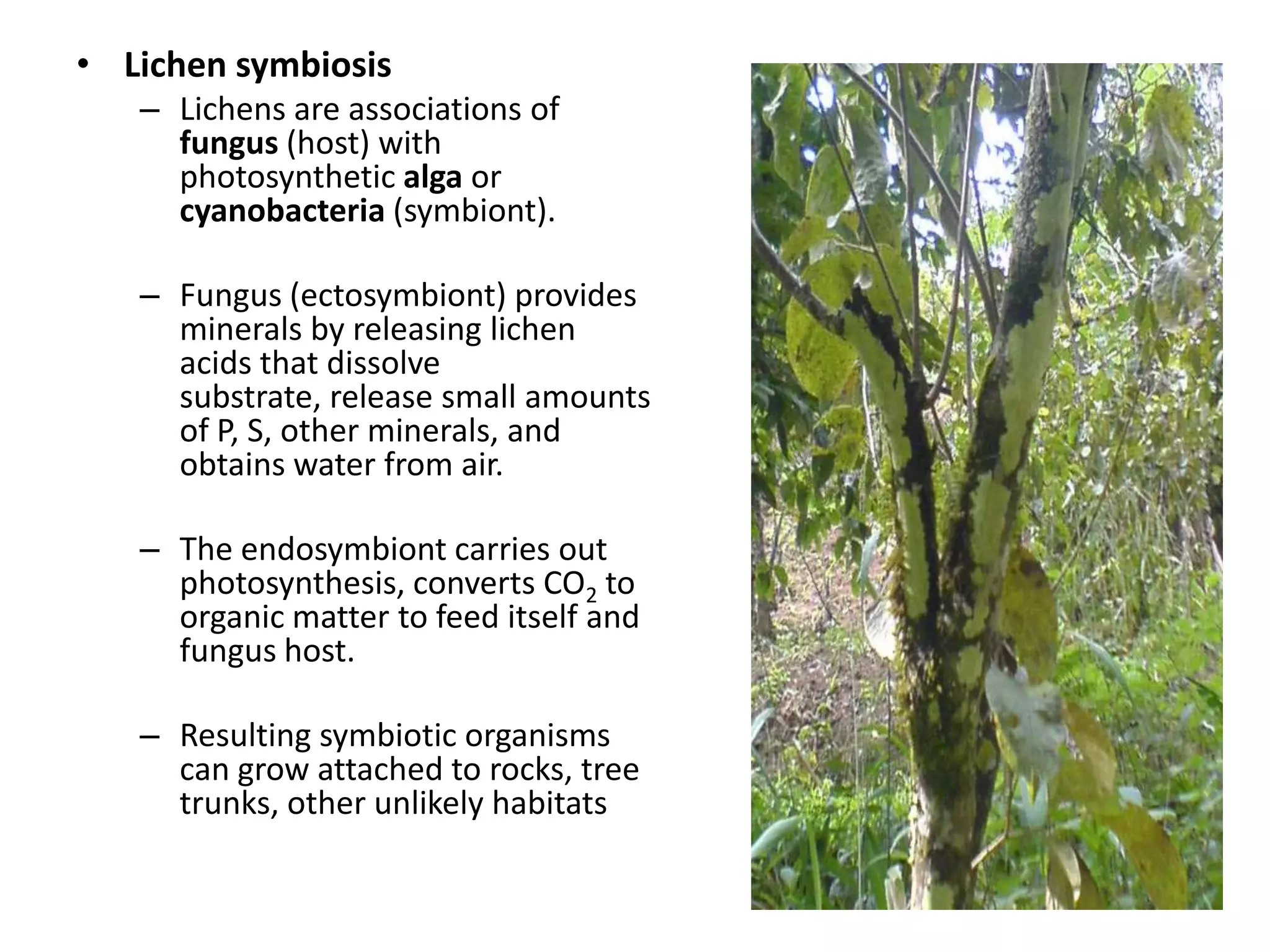
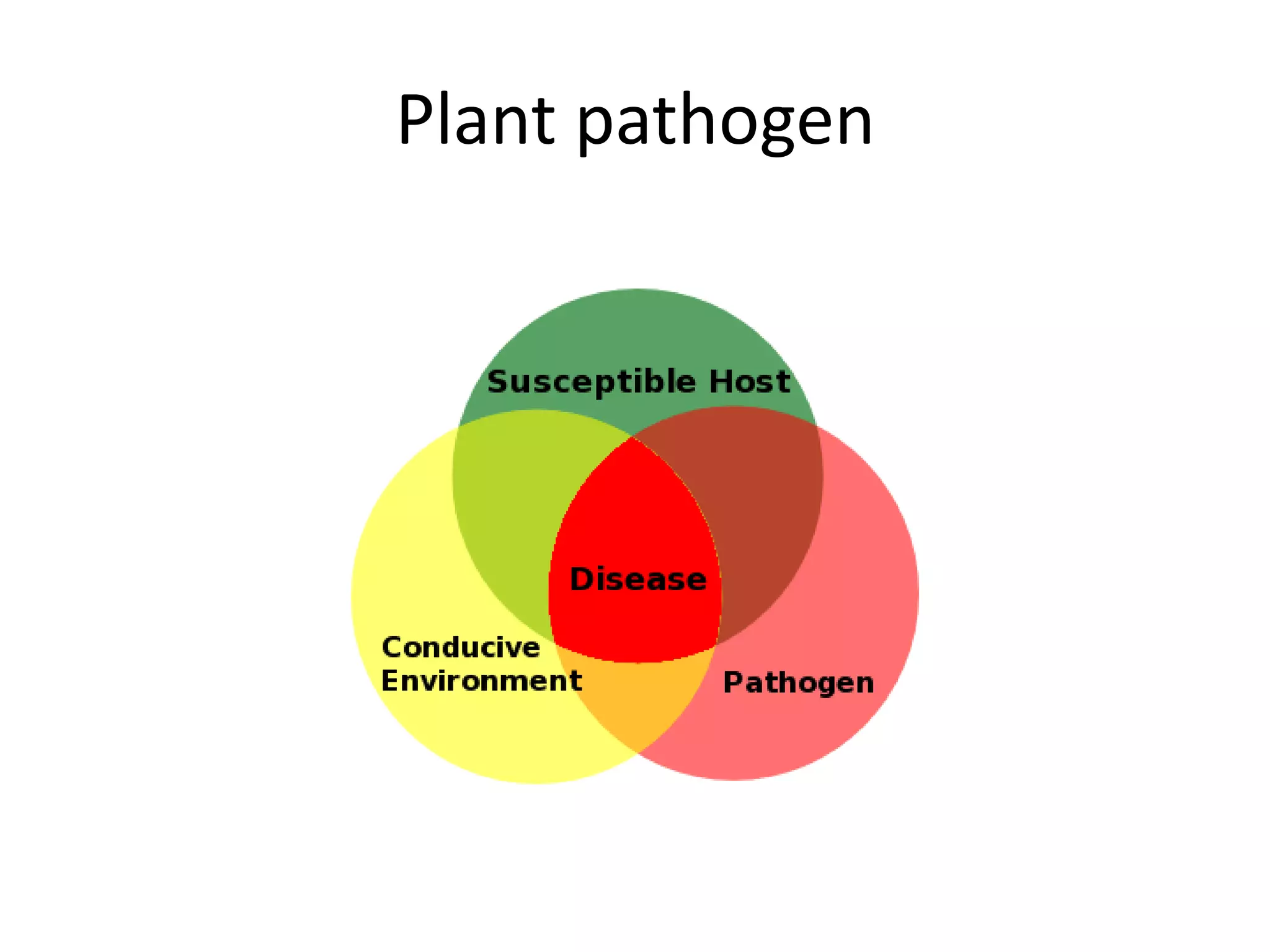
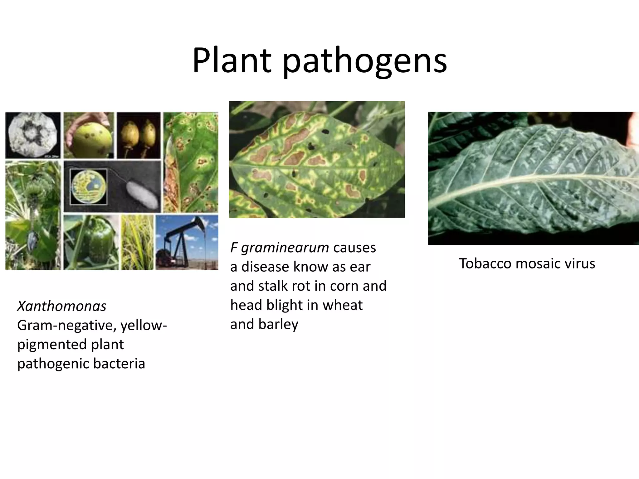
Positive and negative interactions; details on microbial antagonism, lichen symbiosis, and plant pathogens.
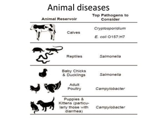
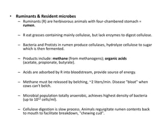
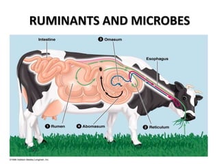
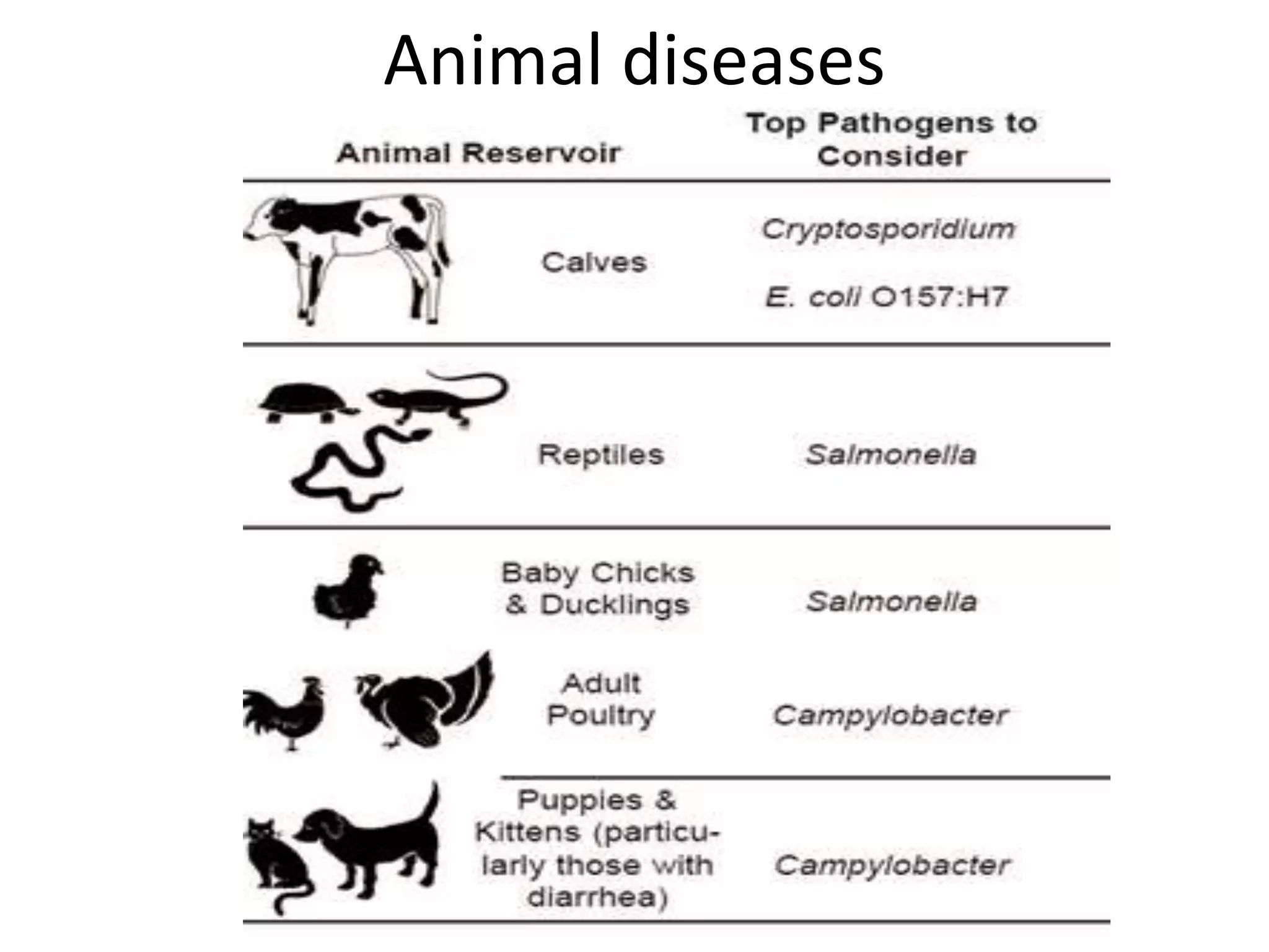
Focus on animal diseases, specifically how ruminants utilize resident microbes to digest cellulose.
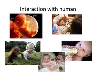
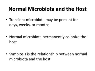
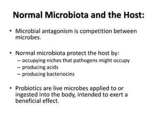
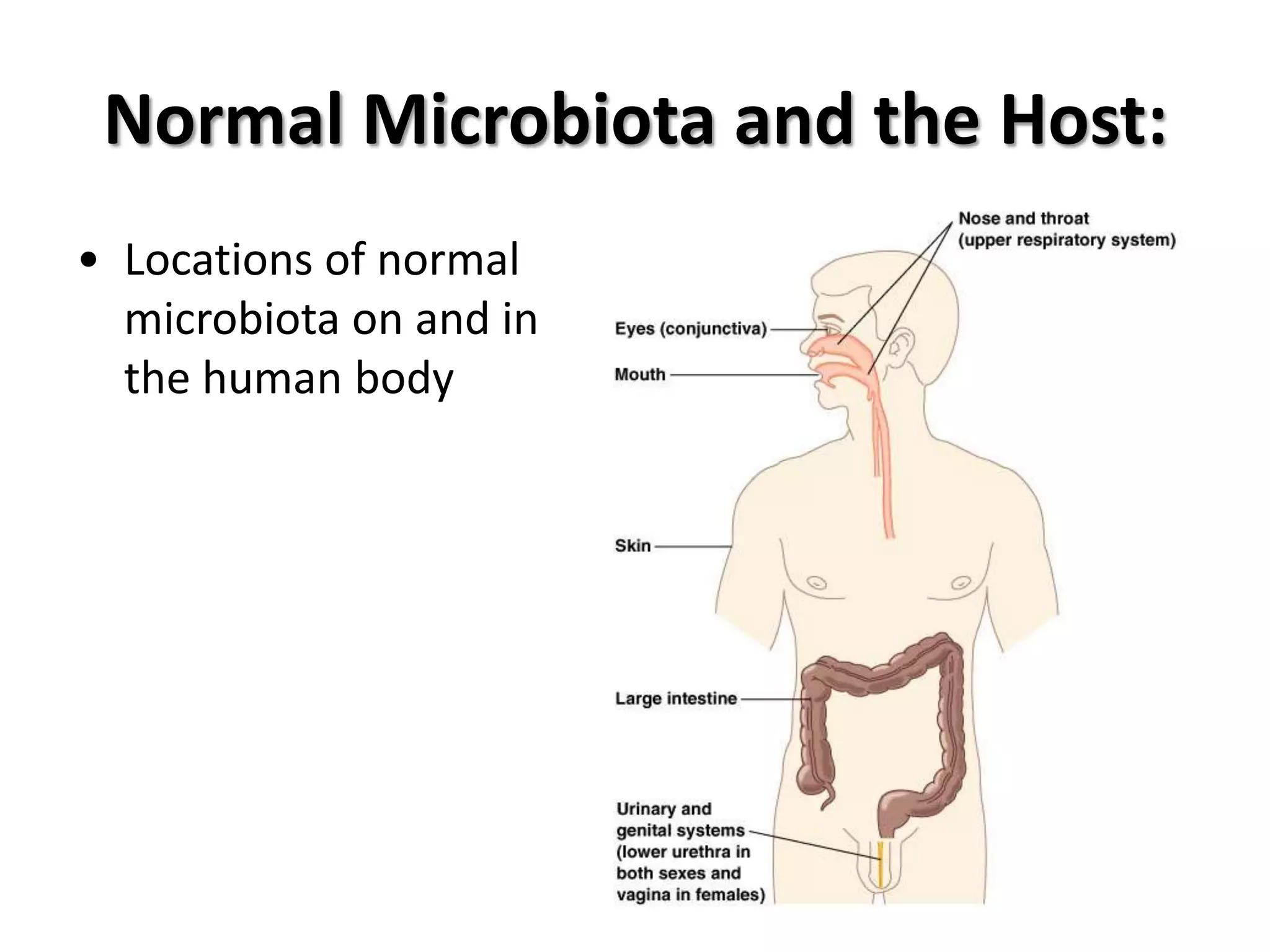
Details of normal microbiota in humans, including transient and permanent colonization and microbial antagonism.
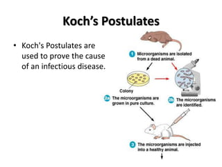
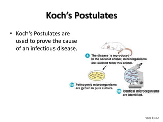
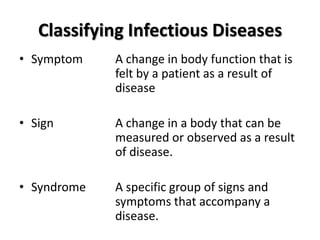
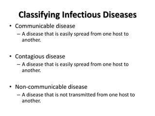
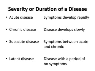
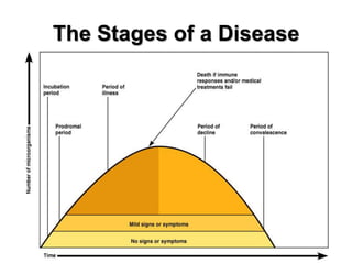
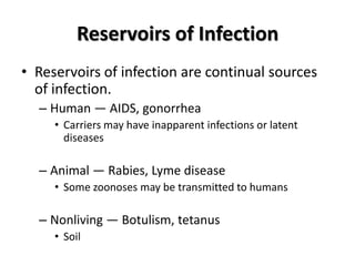
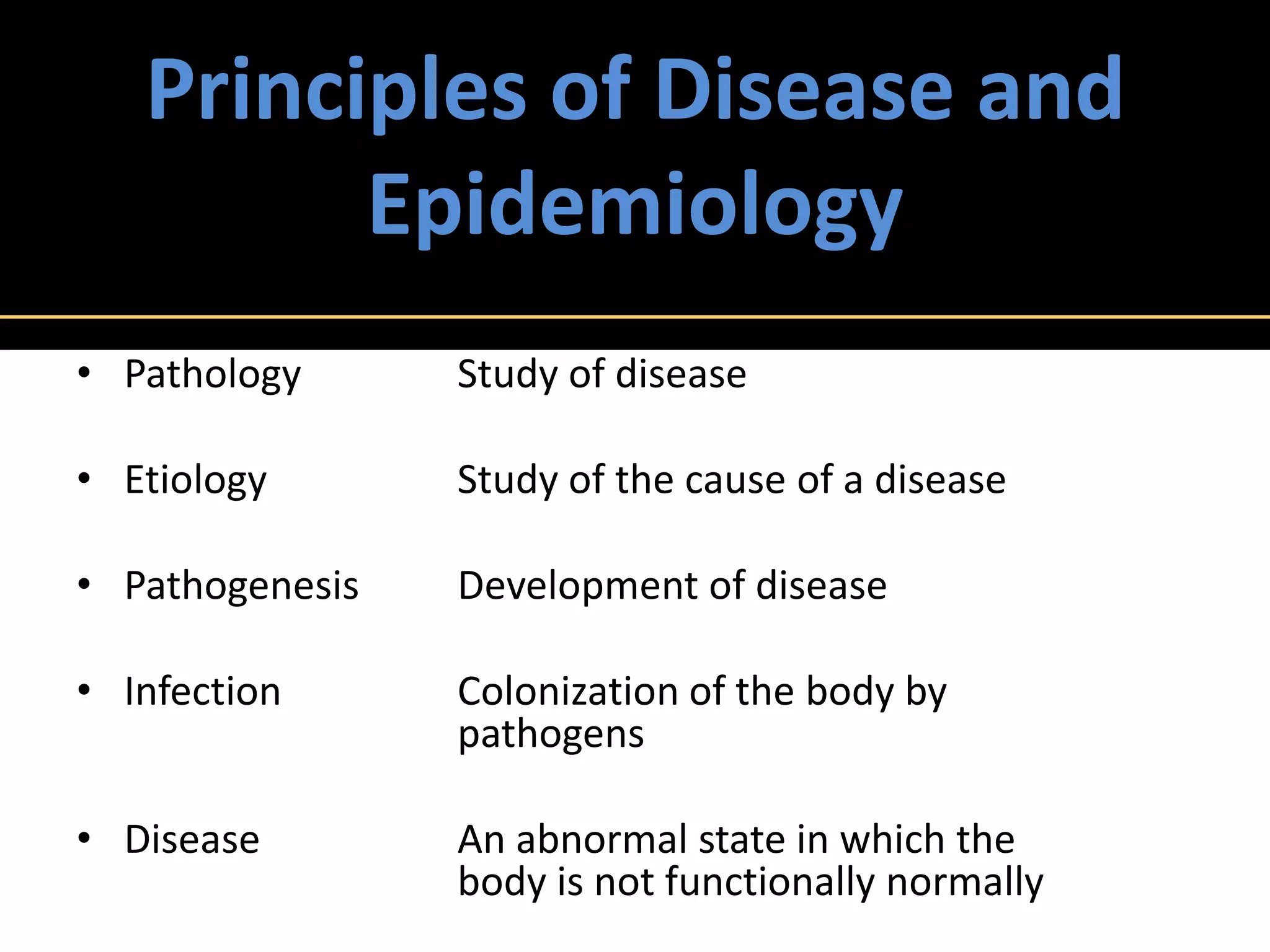
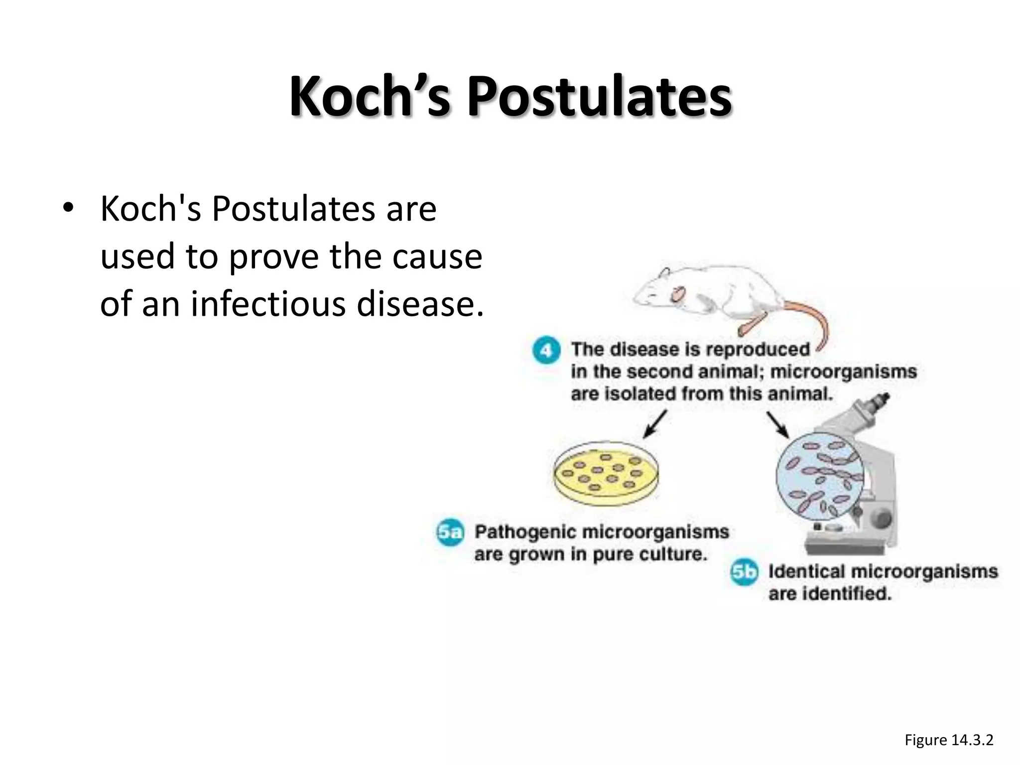
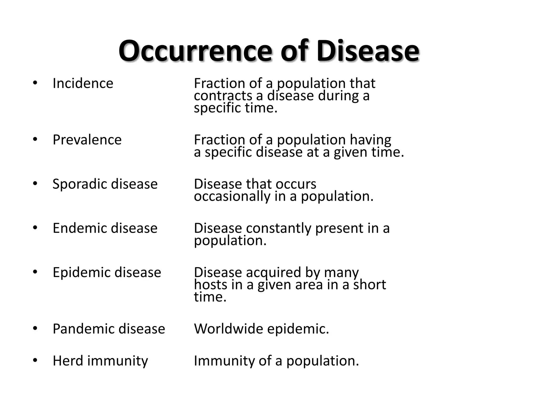
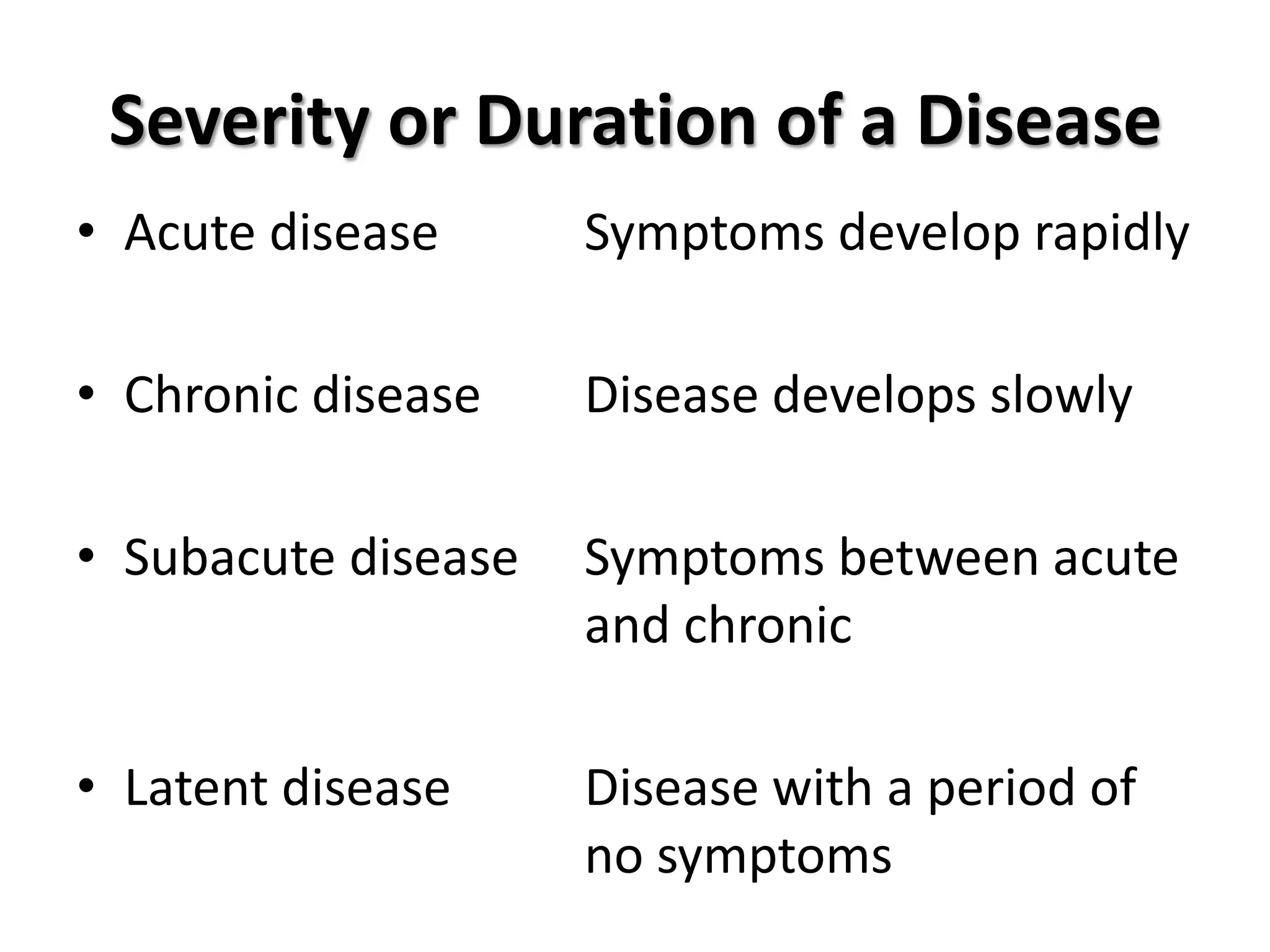
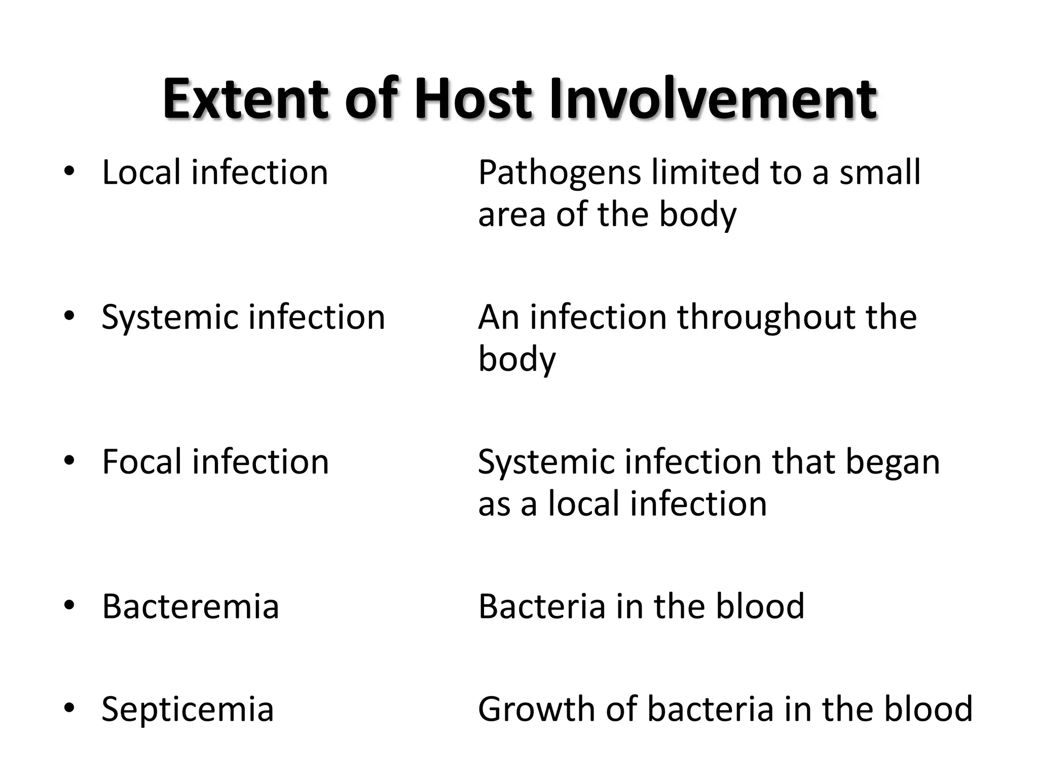
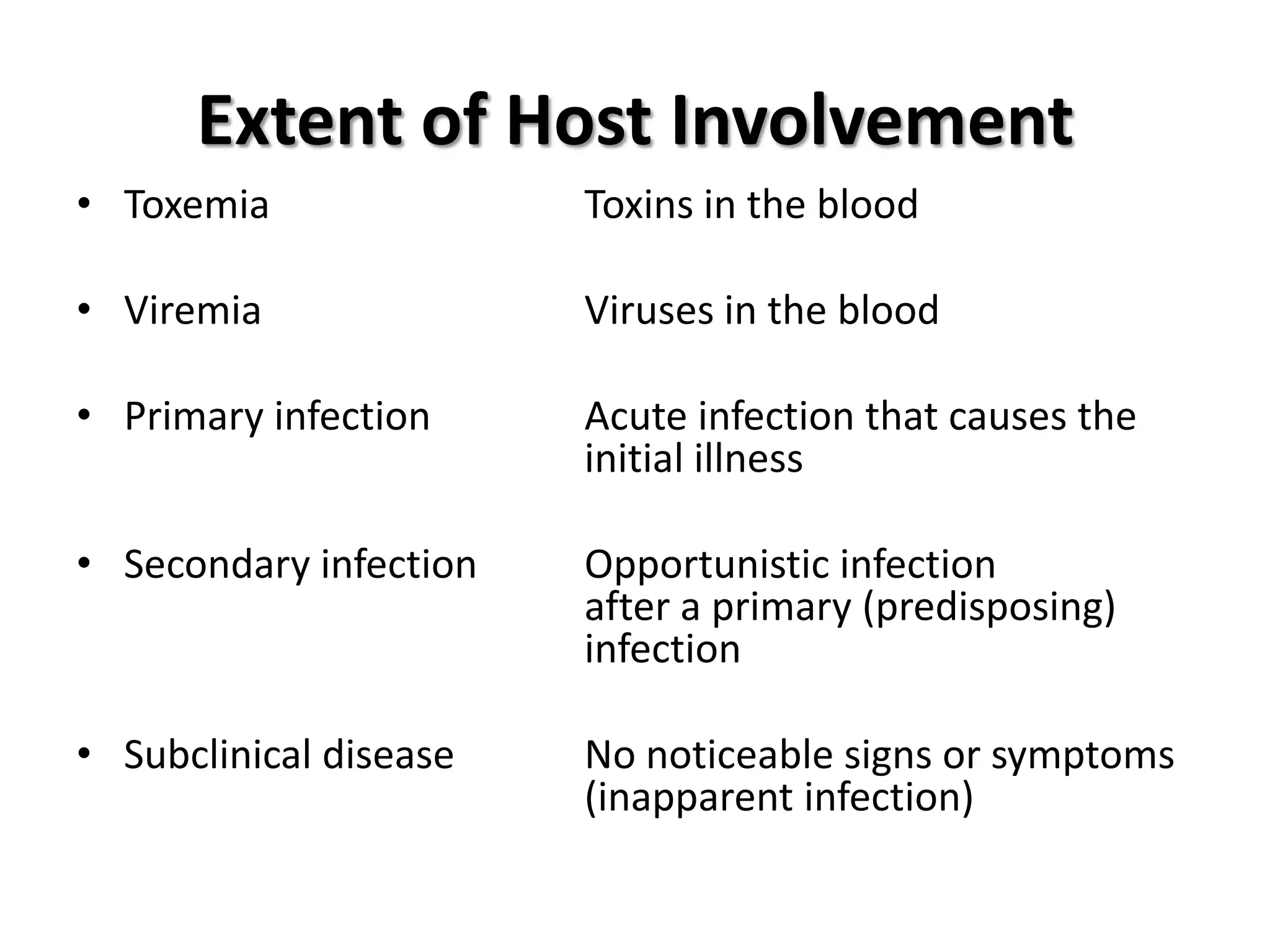
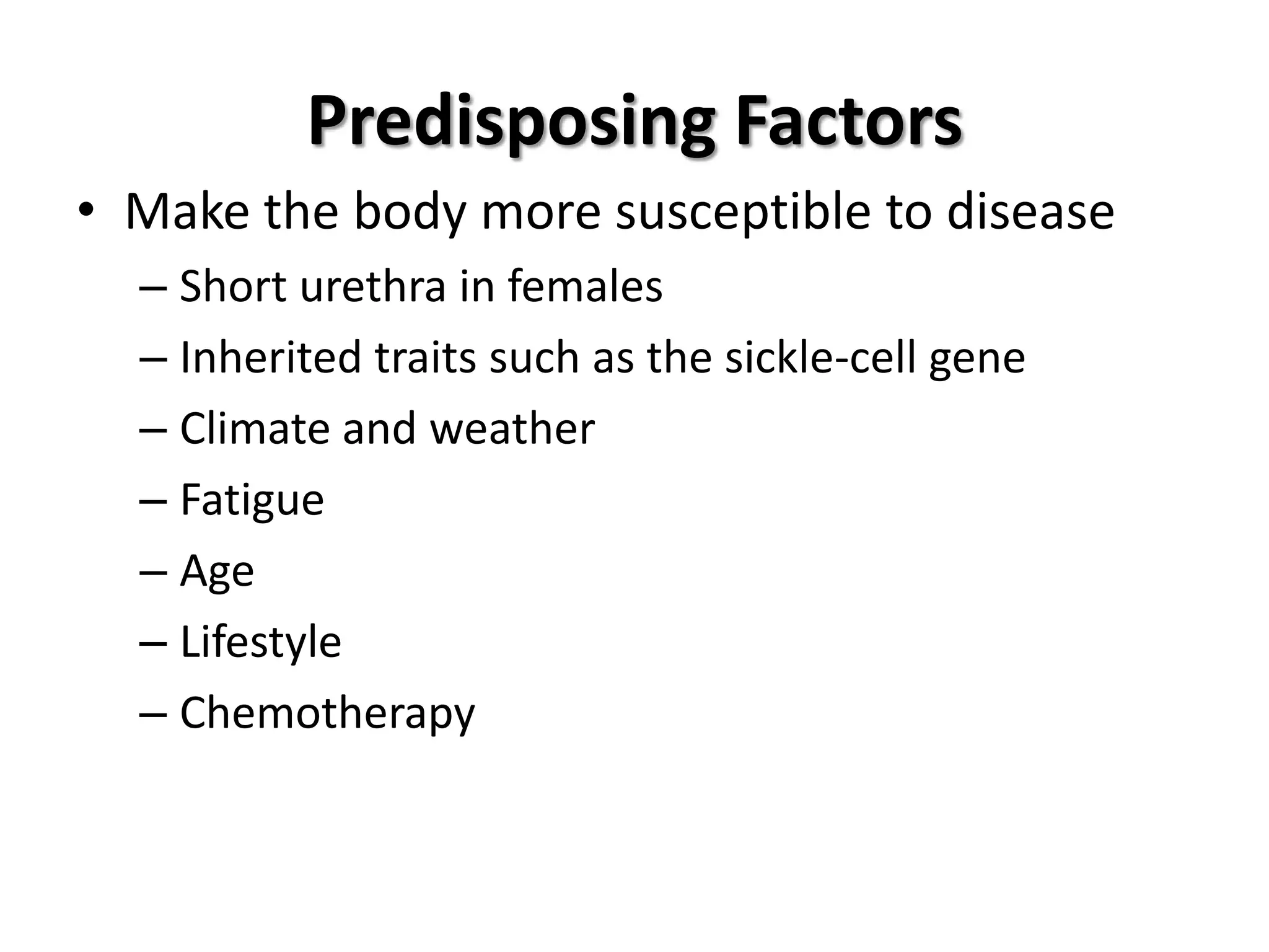
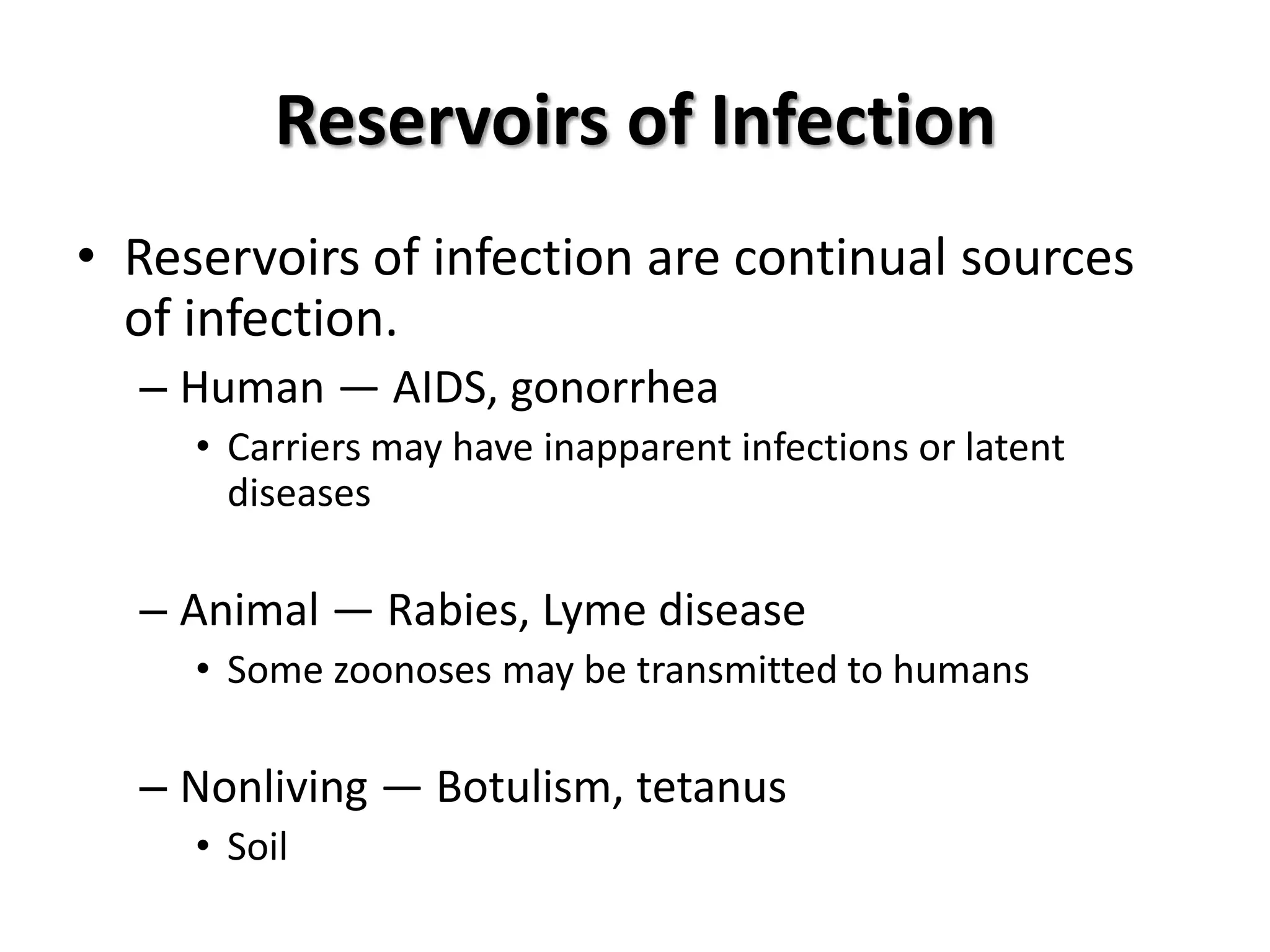
Definitions and principles of disease, including pathology, etiology, infection, and types of diseases. Classifications of infectious diseases based on symptoms, transmission type, and occurrence.
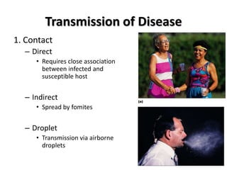
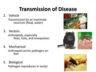
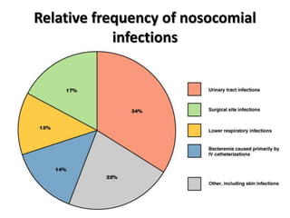
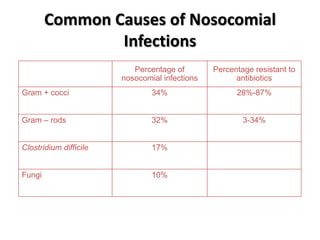
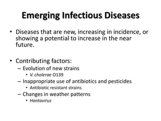
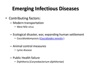
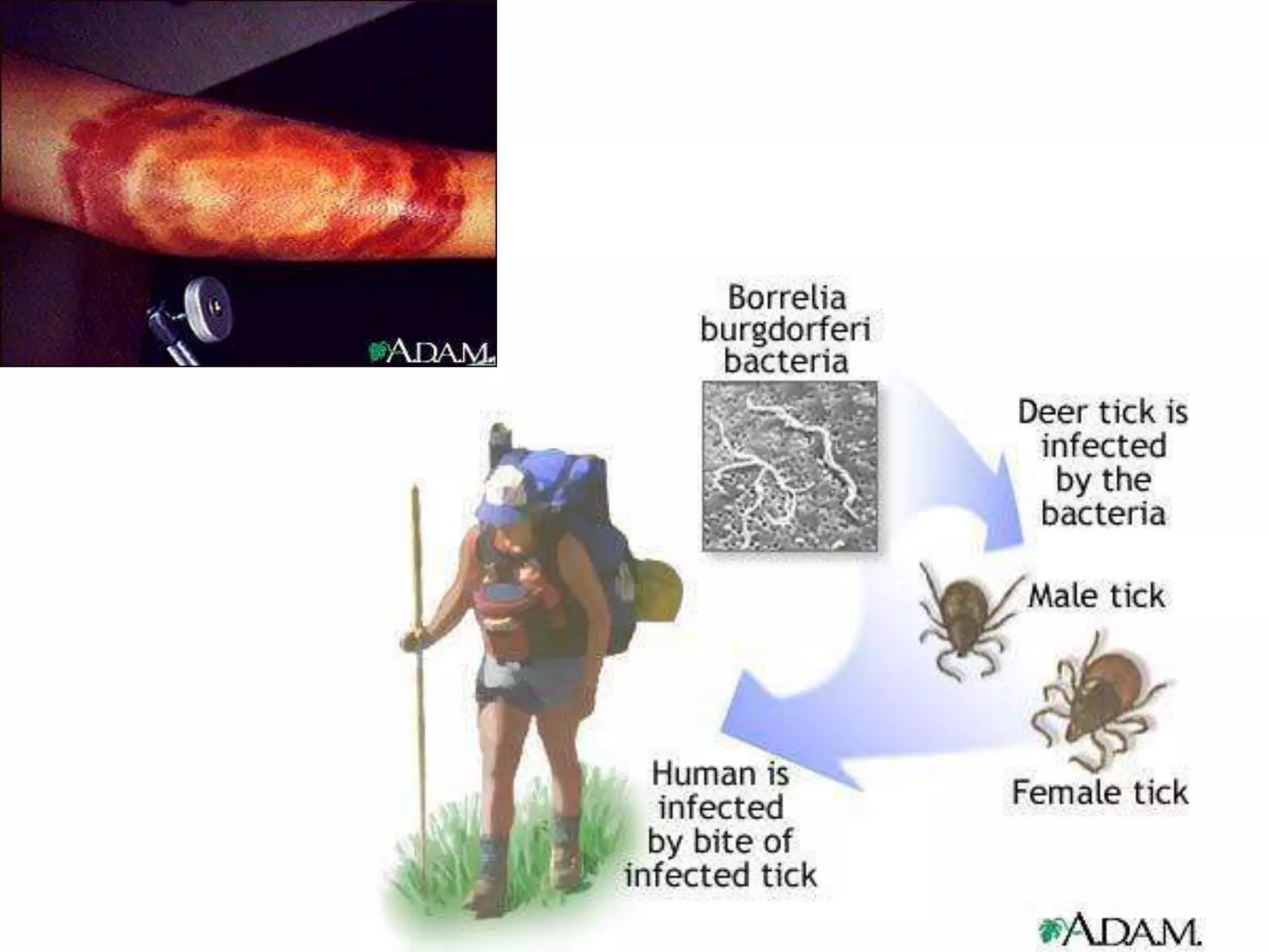
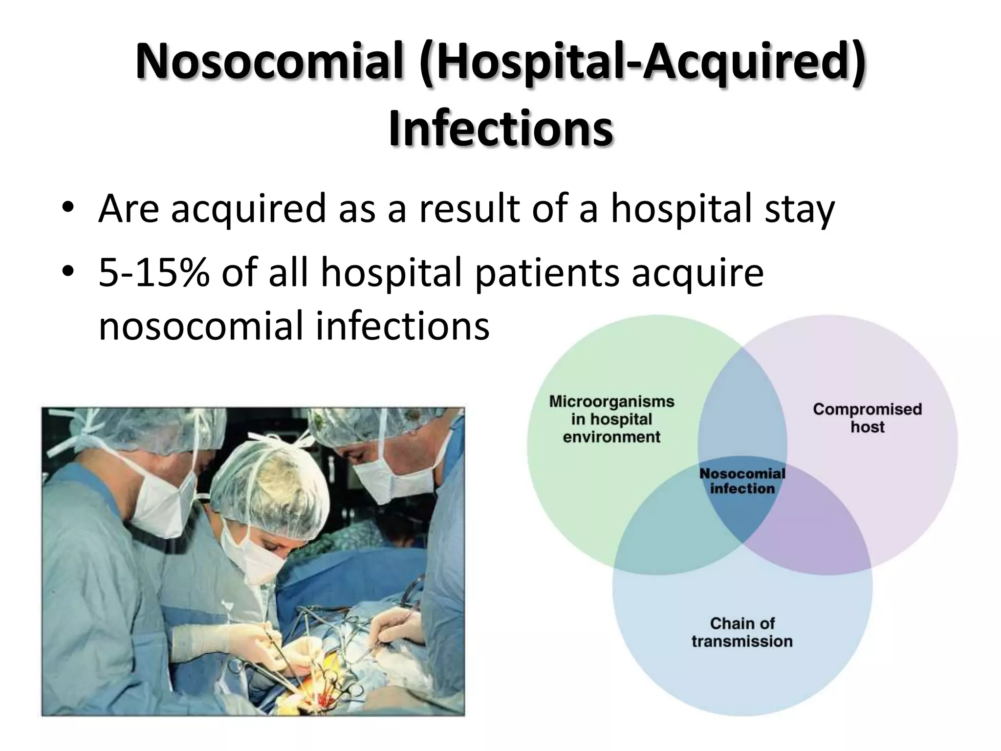
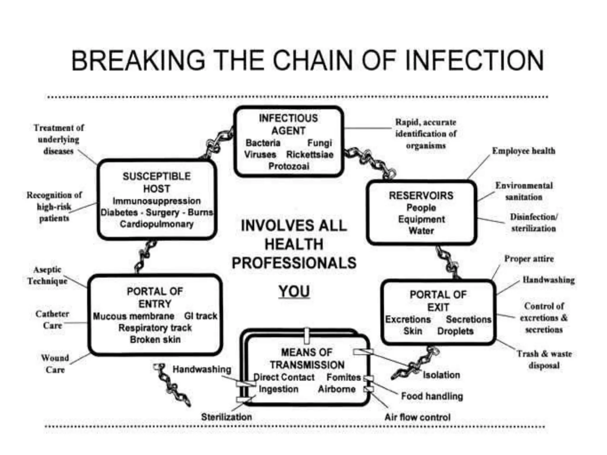
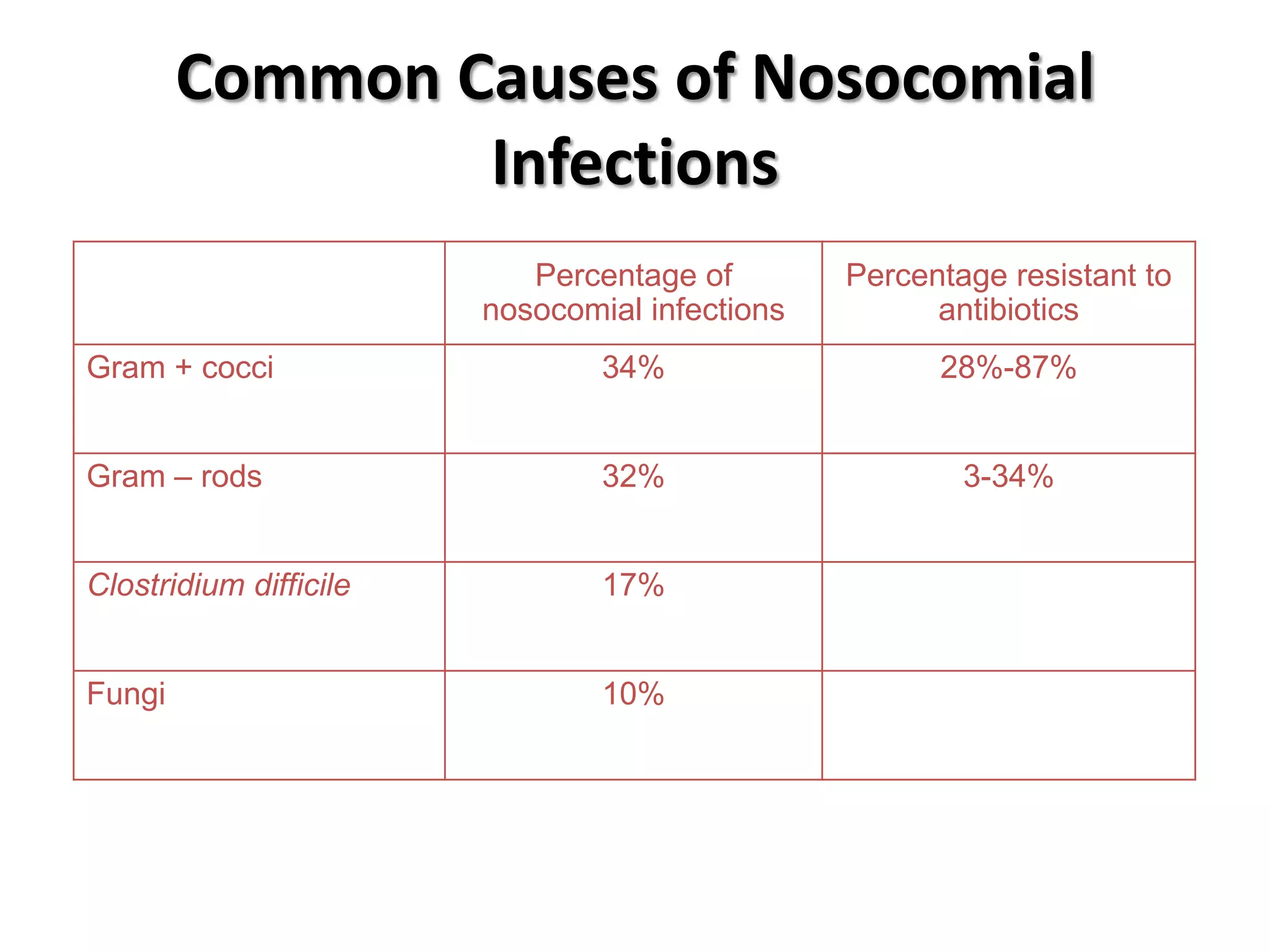
Mechanisms of disease transmission, including direct contact, vectors, and vehicles.Overview of hospital-acquired infections: frequency, causes, and emerging infectious diseases.
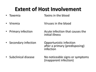
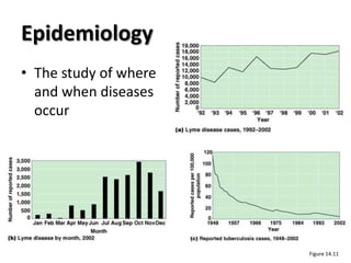
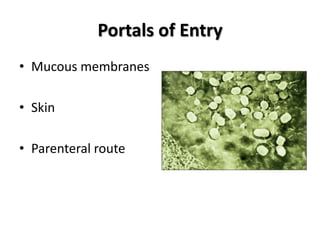
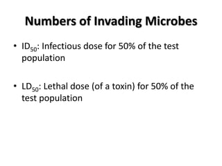
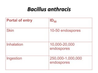
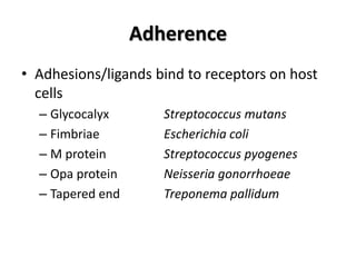
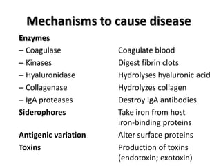
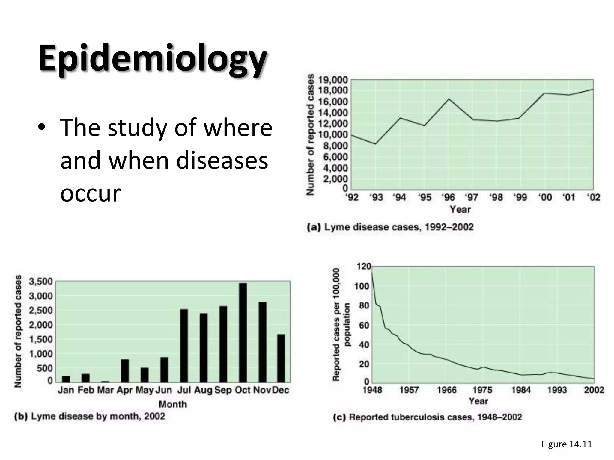
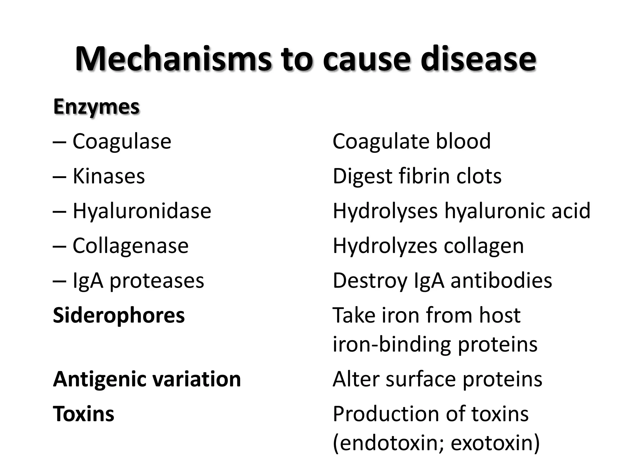
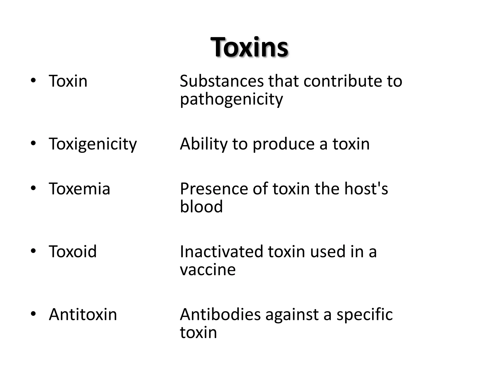
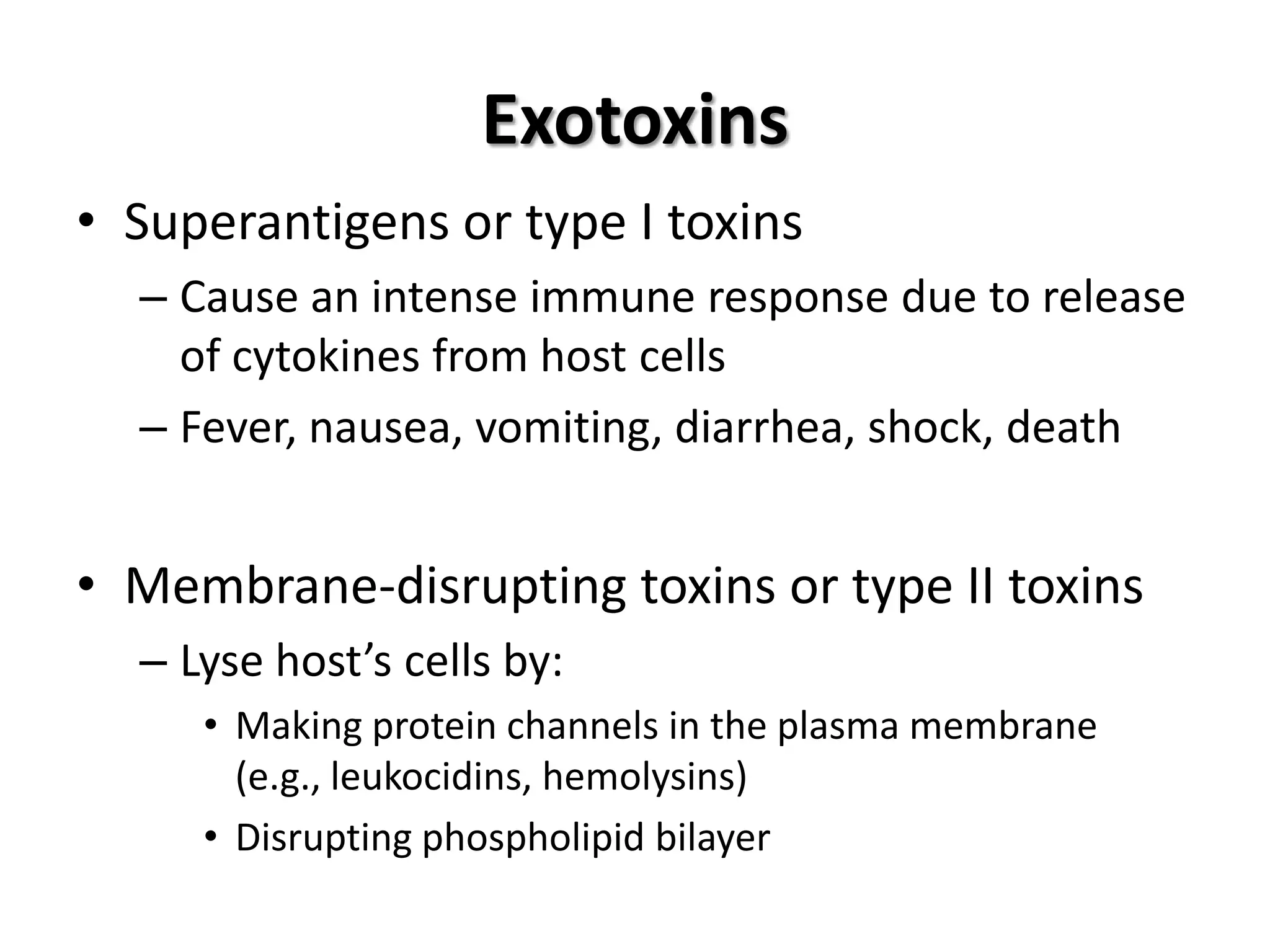
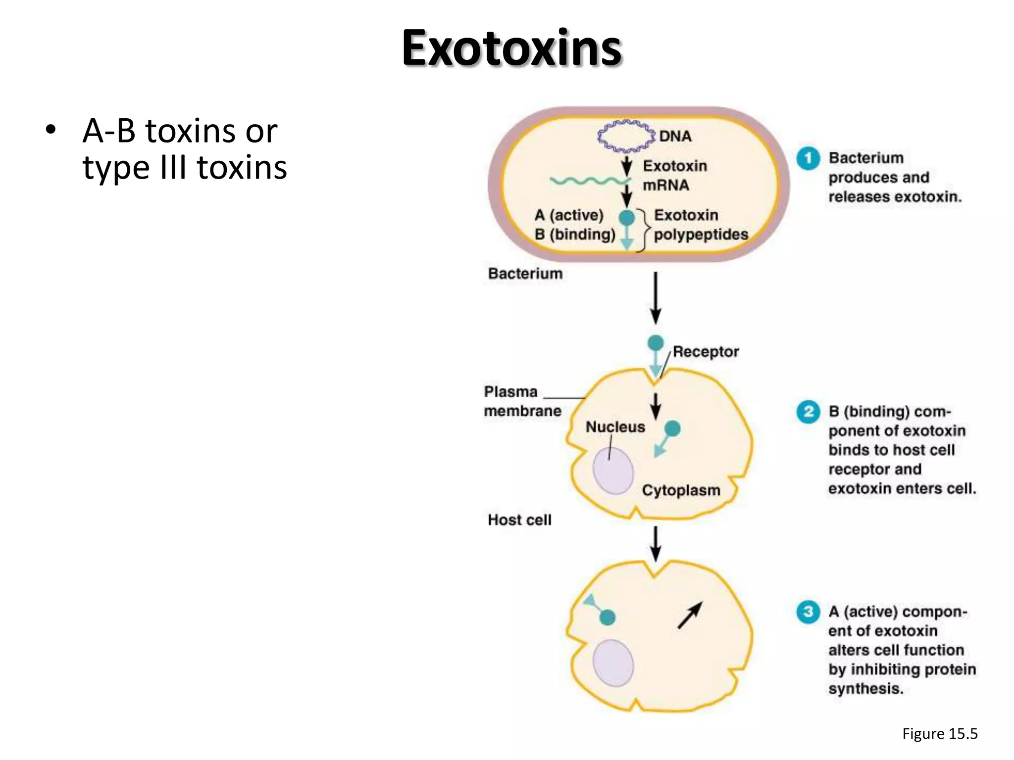
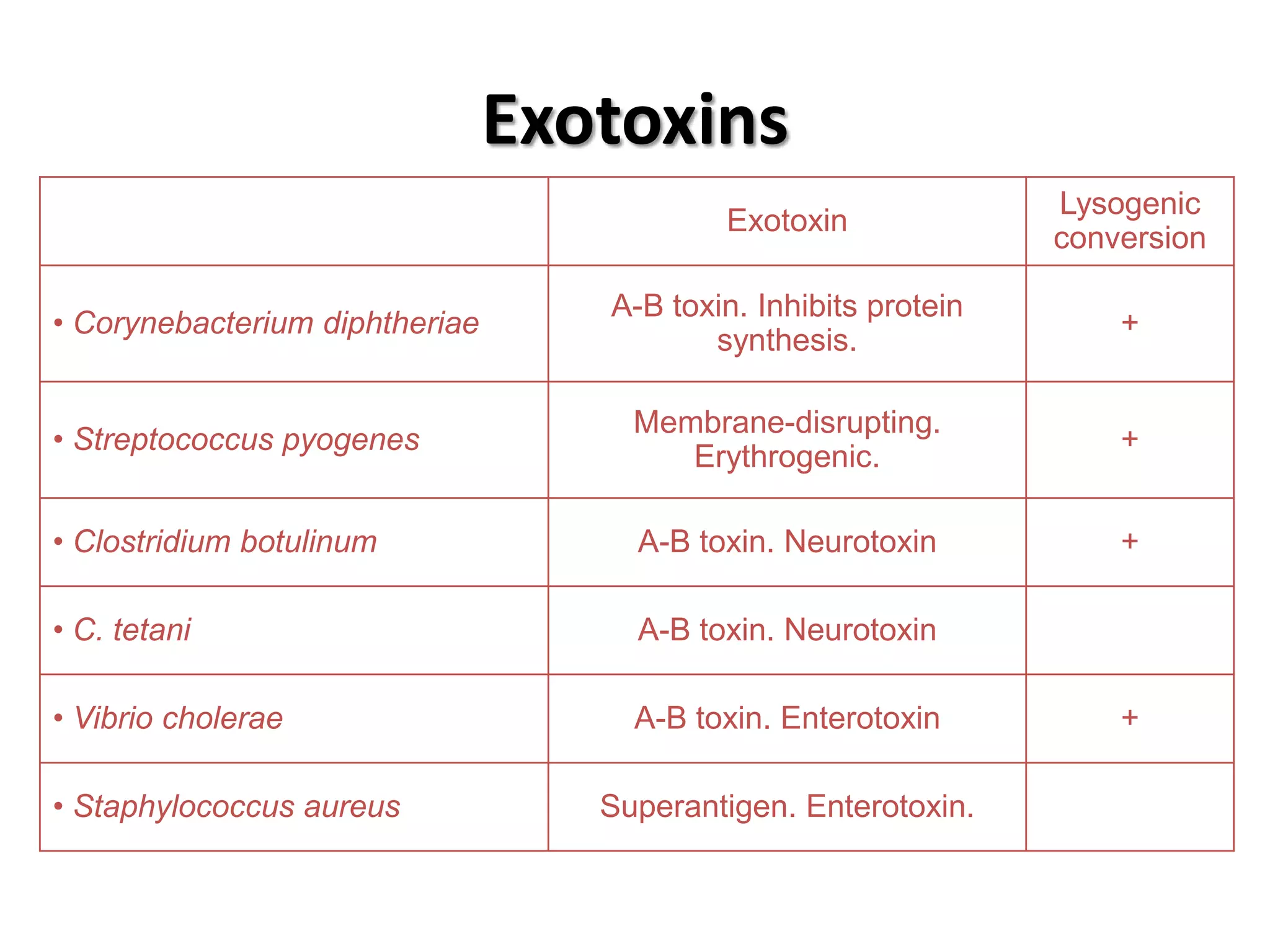
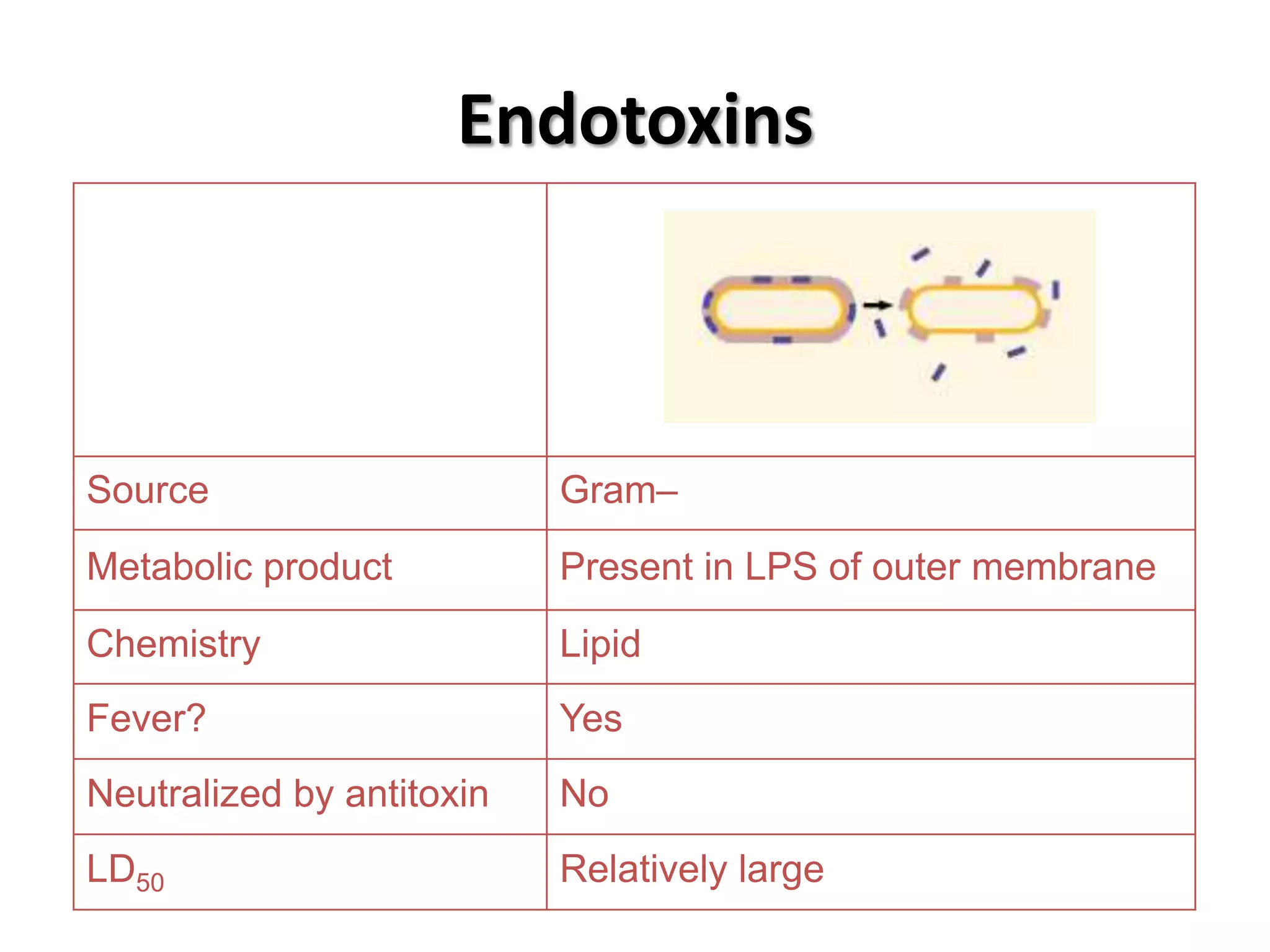
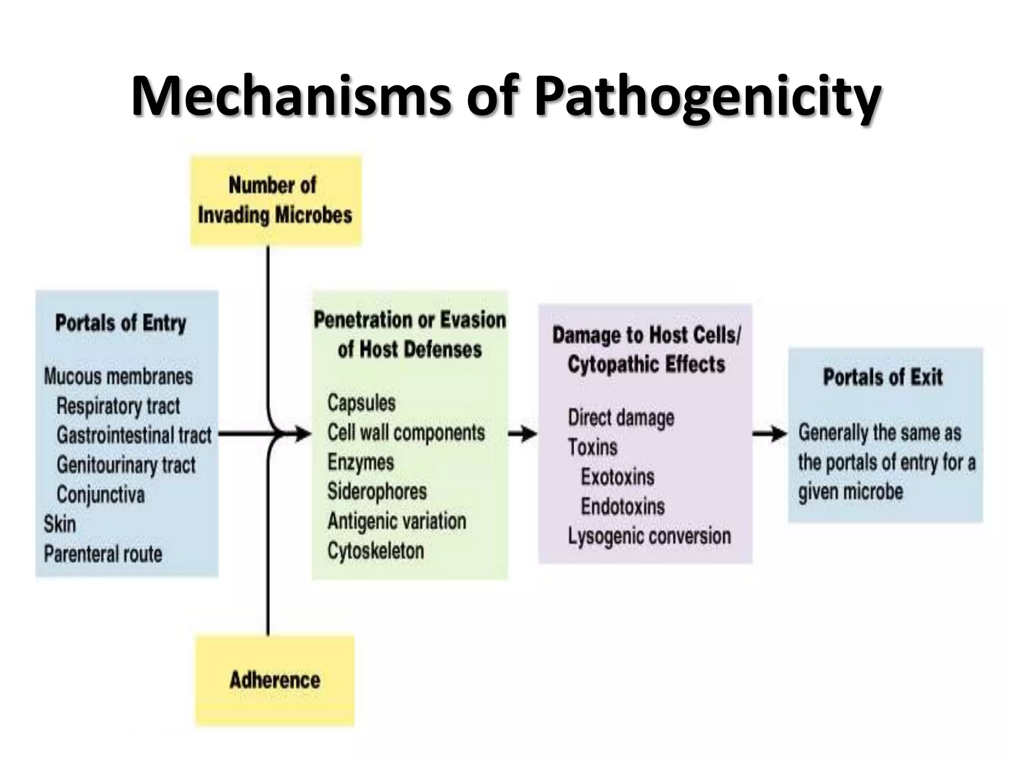
Study of disease occurrence, with a focus on CDC reporting, morbidity, and mortality definitions.Mechanisms of pathogenicity, including portals of entry, infection dose, adherence, and toxins.
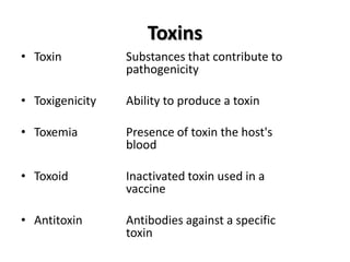
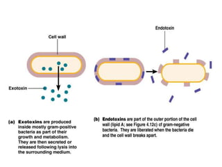
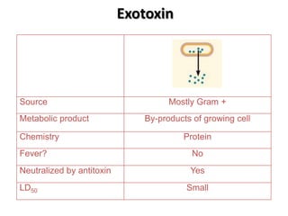
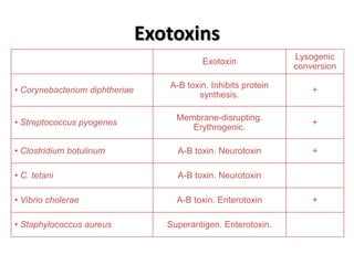
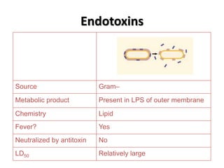
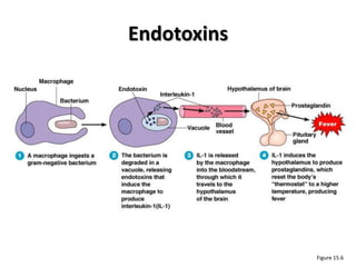
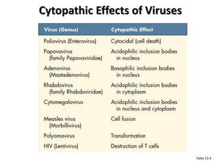
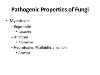
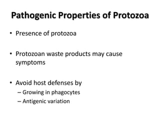
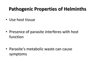
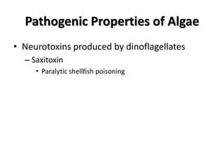
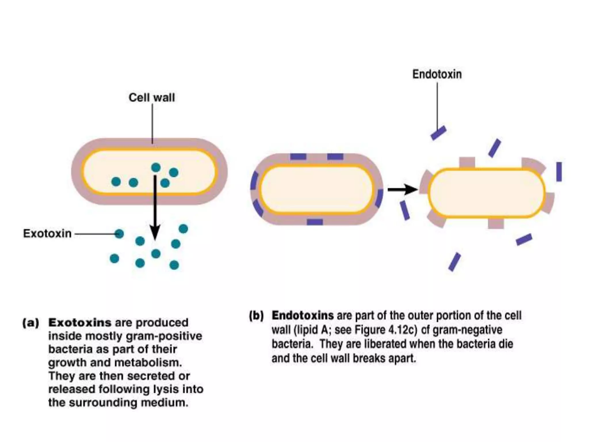
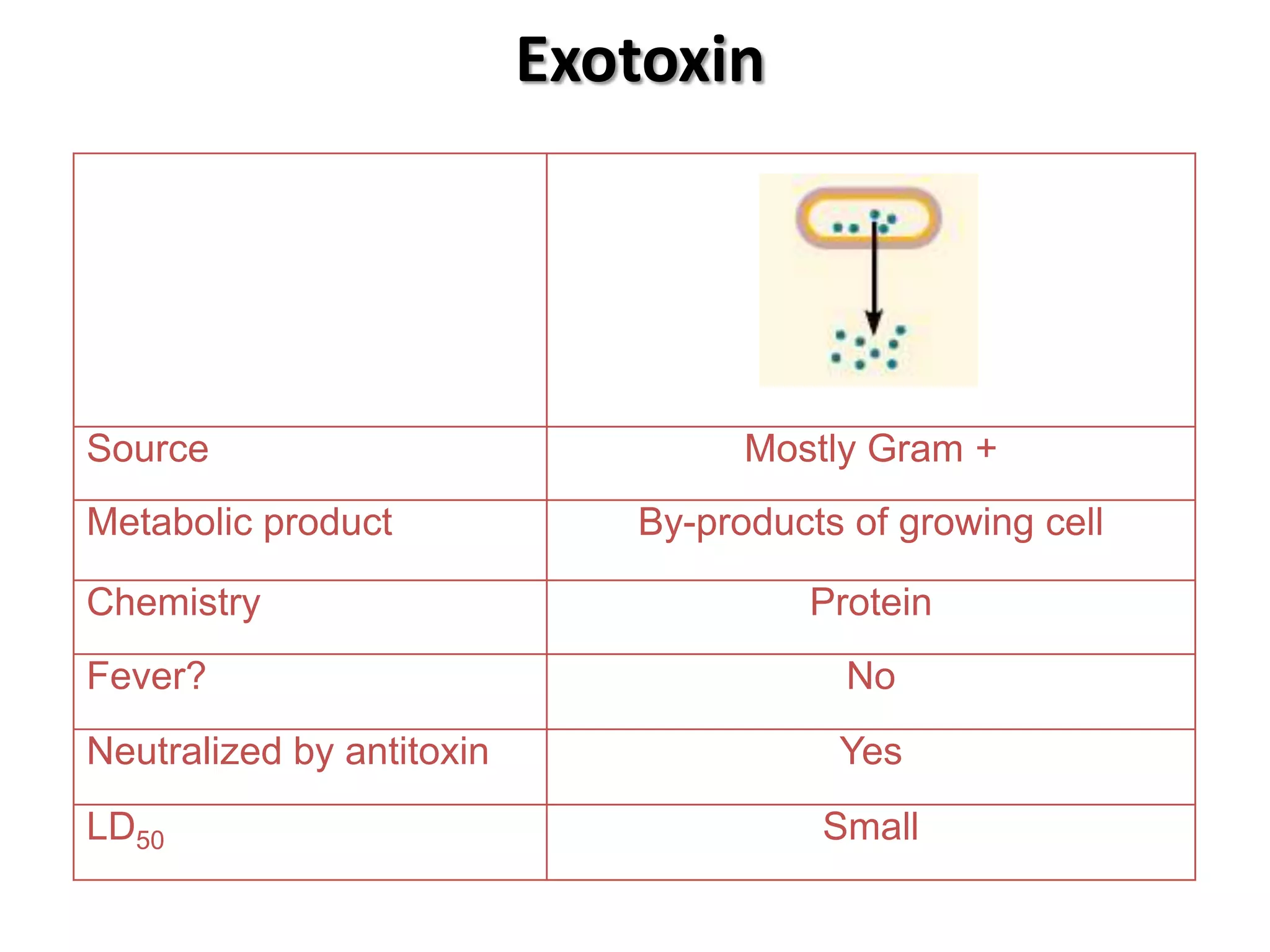
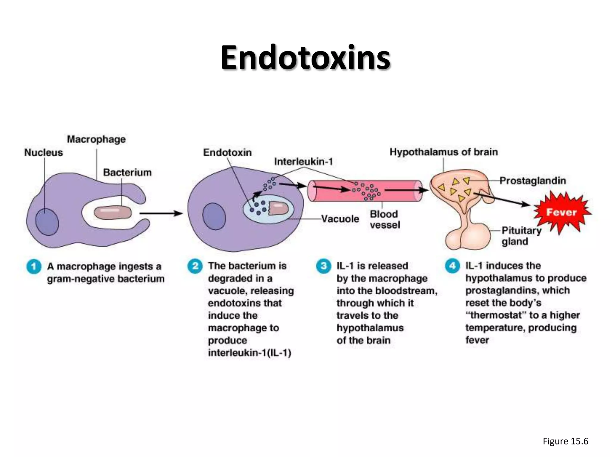
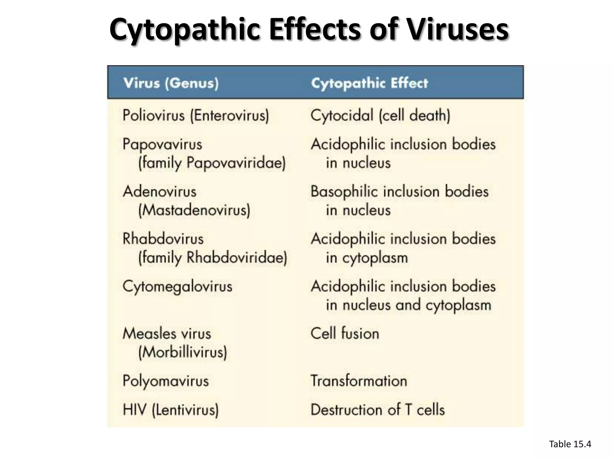
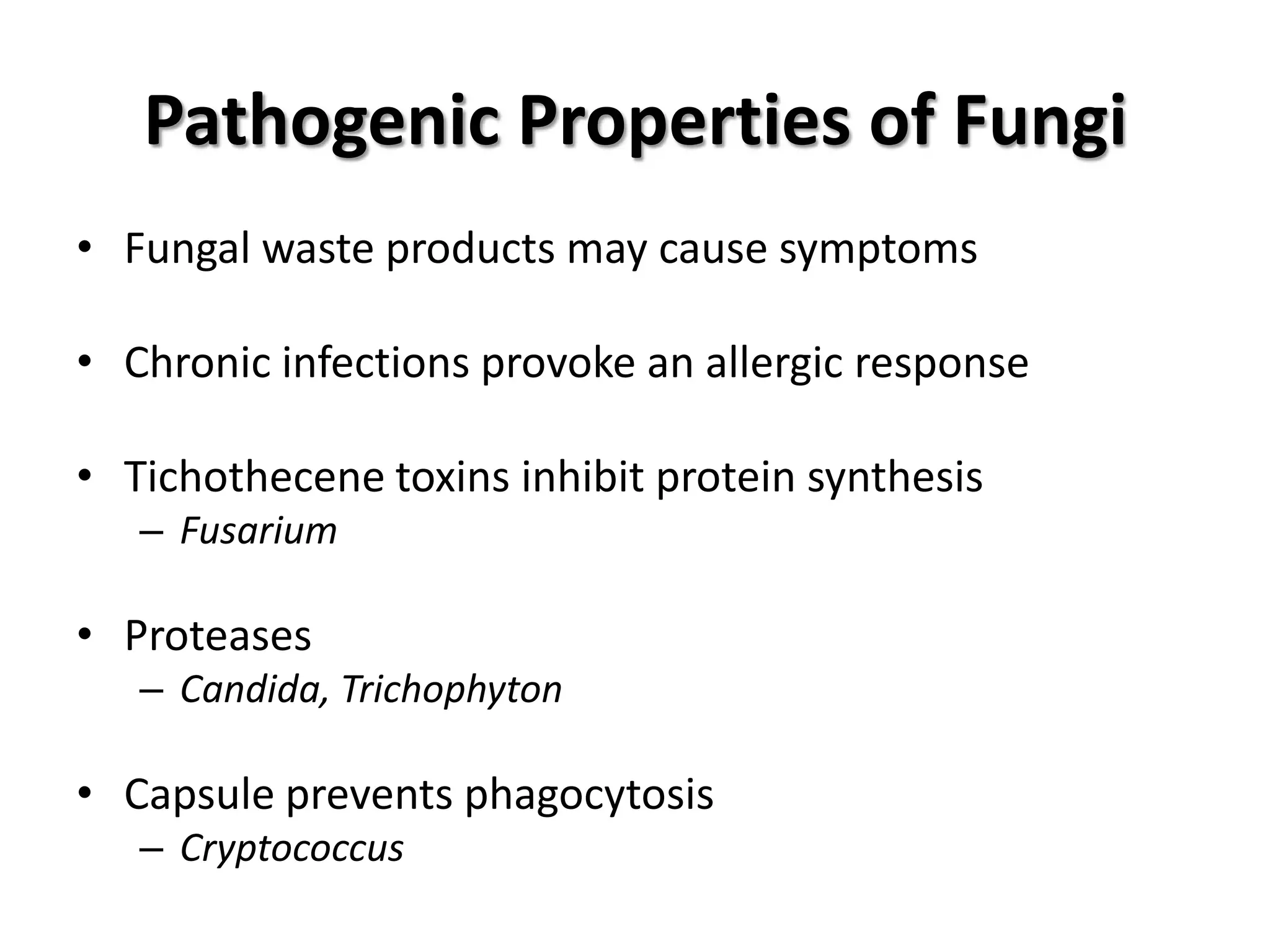
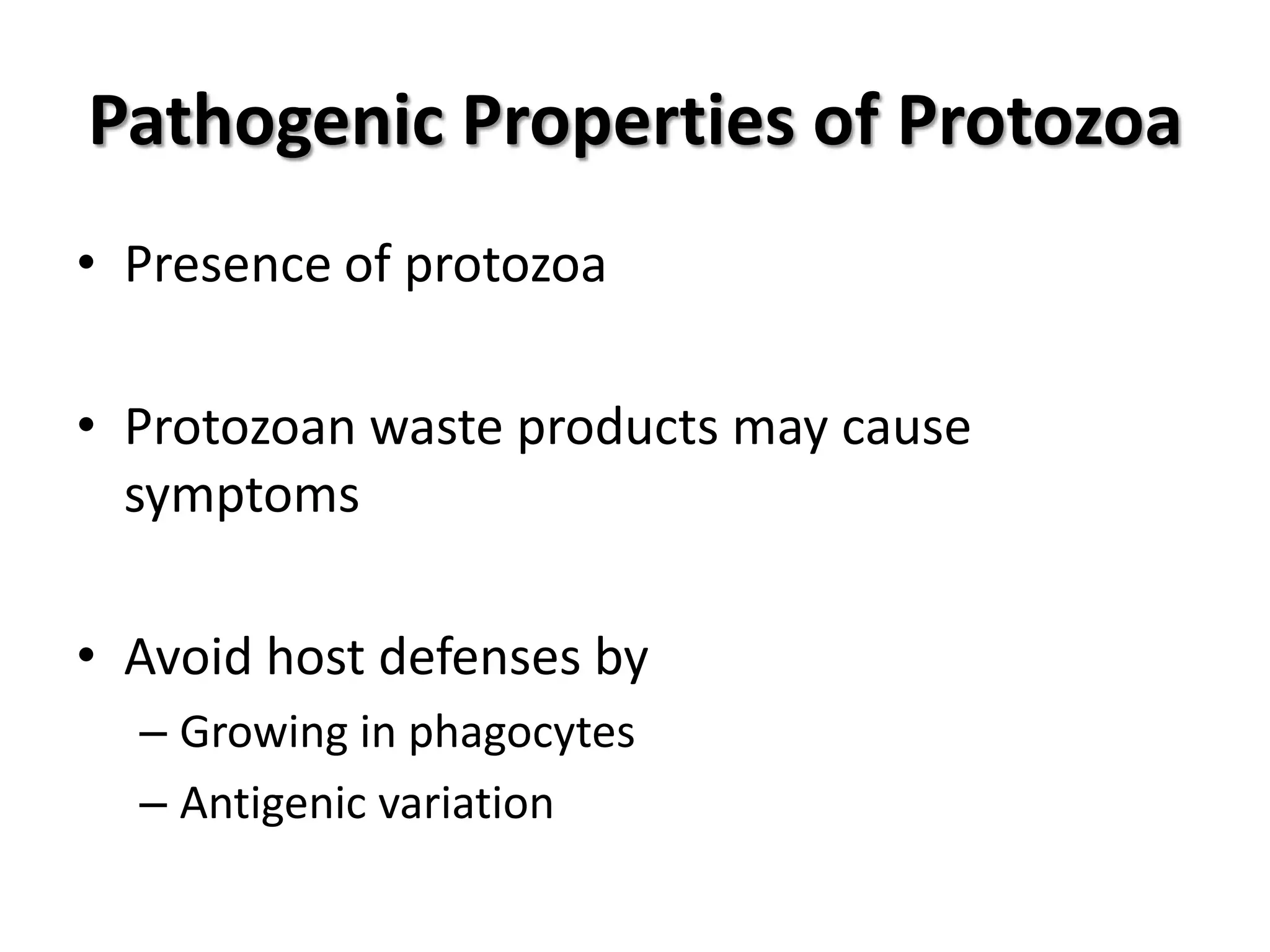
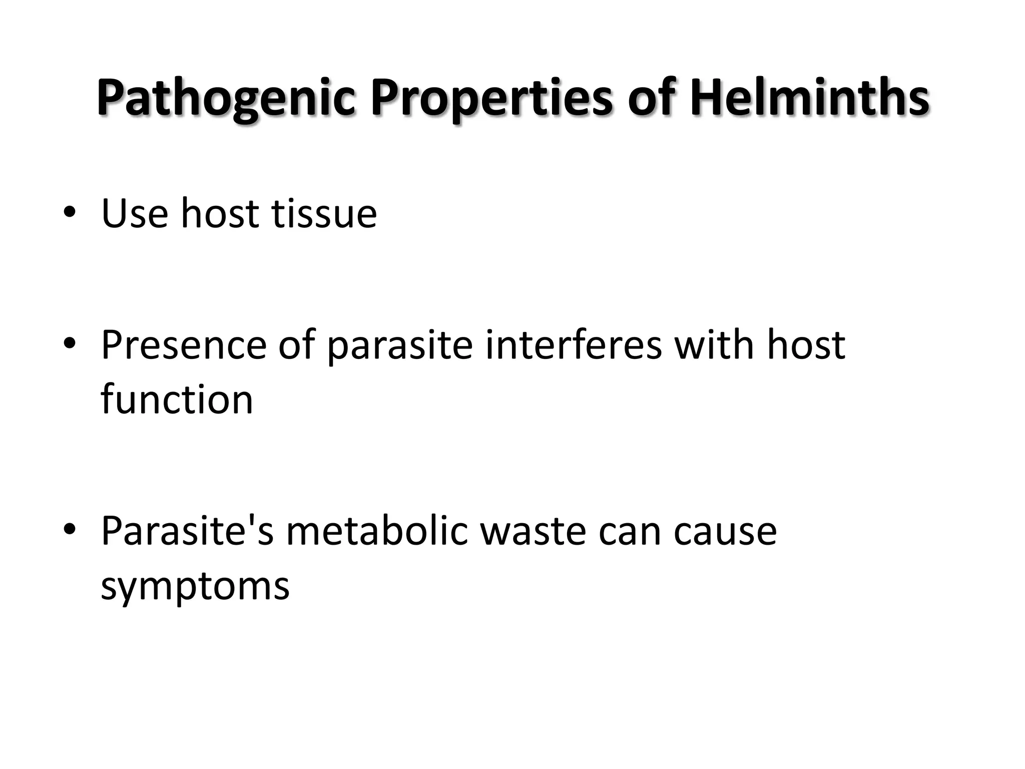
Properties of toxins and pathogens including bacteria, viruses, fungi, protozoa, and helminths.
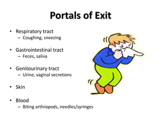
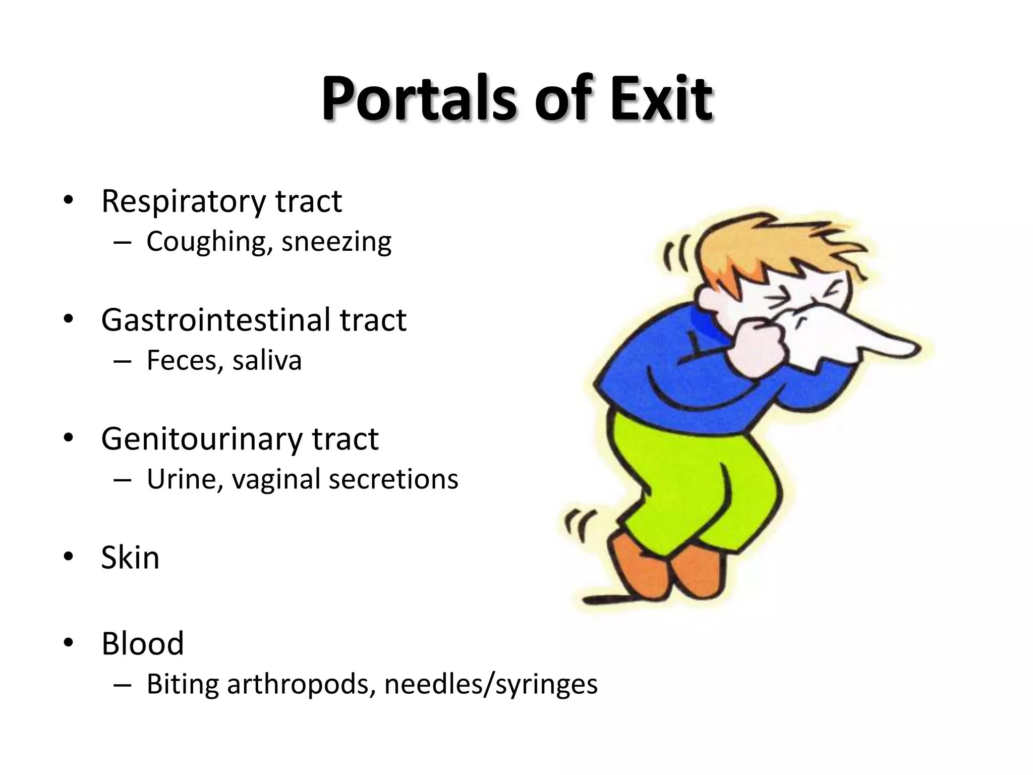
Mechanisms of how pathogens exit the host body via various routes like respiratory and gastrointestinal.


![UiPath Automation Suite Installation (Hands-On) [2/3]](https://cdn.slidesharecdn.com/ss_thumbnails/automationsuitecommunitysession2-251015095633-a6d862f1-thumbnail.jpg?width=600ounds&width=560&fit=bounds)


