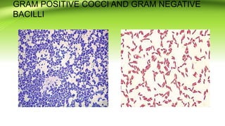
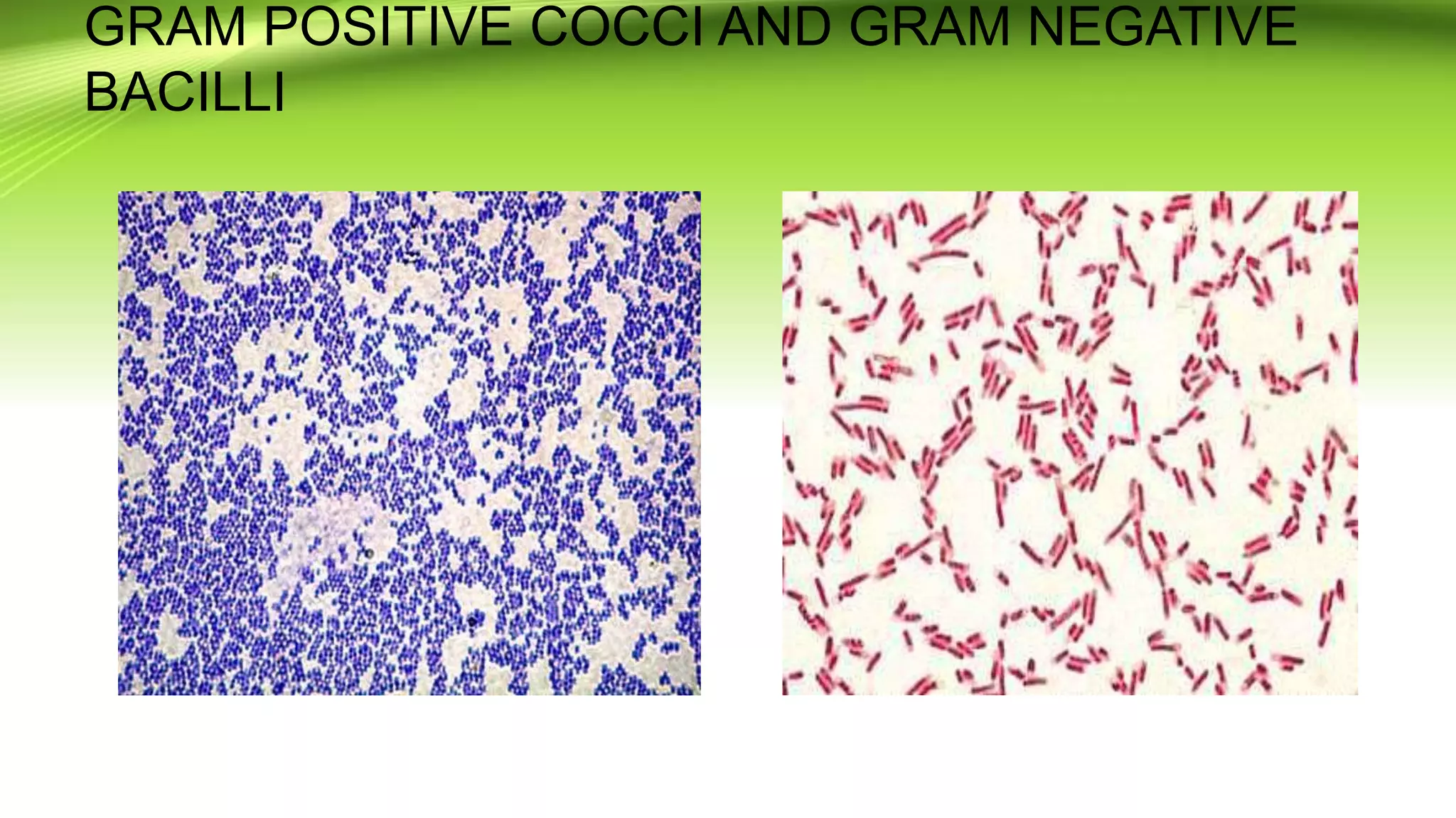
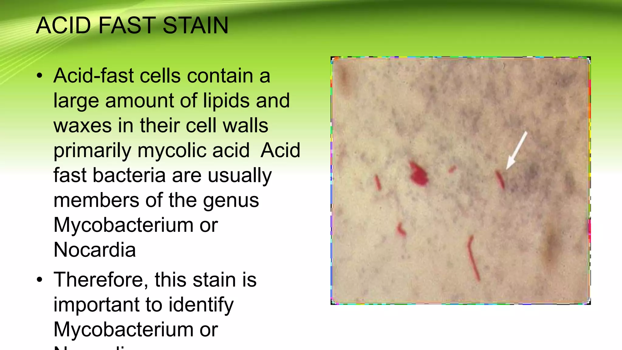
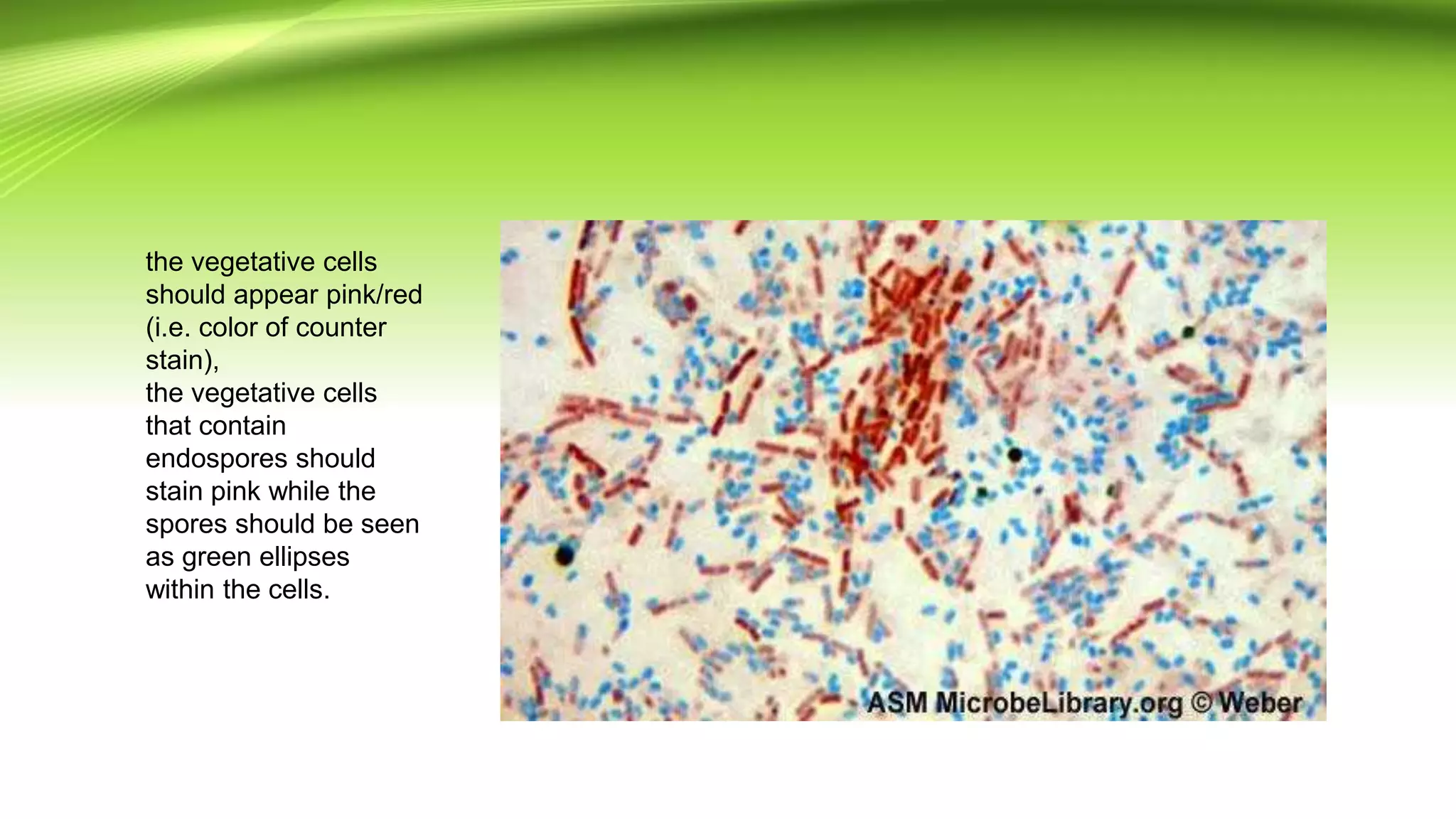
KEMBAR78
STAINING METHODS | PPTX
STAINING METHODS
PRESENTED BY:MAYURI CHELKAR
GUIDED BY:DR.UMA A TUMLAM MAM
GRAM STAINING:PRINCIPLES
• Gram staining is used to determine gram status to classify
bacteria broadly. It is based on thecomposition of their
cell wall. Gram staining uses crystal violet to stain cell
walls, iodine as a mordant,and a fuchsin or safranin
counterstain to mark all bacteria. Gram status is important
in medicine; the presence or absence of a cell wall will
change the bacterium's susceptibility to some antibiotics.
Gram-positive bacteria stain dark blue or violet.
• Their cell wall is typically rich with peptidoglycan and
lacks the secondary membrane and lipopolysaccharide
layer found in Gram-negative bacteria
STEPS
• Crystal violet acts as the primary stain. Crystal violet mayalso
be used as a simple stain because it dyes the cell wallof any
bacteria.
• 2. Gram’s iodine acts as a mordant (Helps to fix the primary
dye to the cell wall).
• 3. Decolorizer is used next to remove the primary stain (crystal
violet) from Gram Negative bacteria (those with LPS imbedded
in their cell walls). Decolorizer is composed of an organic
solvent, such as, acetone or ethanol or a combination of both.)
• 4. Finally, a counter stain (Safranin), is applied to stain those
cells (Gram Negative) that have lost the primary stain as a
result of decolorization
STRUCTURE
GRAM POSITIVE COCCI AND GRAM NEGATIVE
BACILLI
GRAM POSITIVE
• Gram-positive bacteria are those that are stained dark
blue or violet by Gram staining. This is in contrast to
Gram-negative bacteria, which cannot retain the crystal
violet stain, instead taking up the counter stain (safranin
or fuchsine) and appearing red or pink. Grampositive
organisms are able to retain the crystal violet stain
because of the high amount of peptidoglycan in the cell
wall. Gram-positive cell walls typically lack the outer
membrane found in Gram-negative bacteria.
GRAM NEGATIVE BACTERIA
• Gram-negative bacteria are those bacteria that
• do not retain crystal violet dye in the Gram
• staining protocol. In a Gram stain test, a counter
• stain (commonly safranin) is added after the crystal
• violet, coloring all Gram-negative bacteria with a red
• or pink color. The test itself is useful in classifying
• two distinct types of bacteria based on the structural
• differences of their cell walls. On the other
• hand, Gram-positive bacteria will retain the crystal
• violet dye when washed in a decolorizing solution.
• On most Gram-stained
• preparations, Gram-negative
• organisms will appear red or
• pink because they are
• counterstained. Due to
• presence of higher lipid
• content, after alcoholtreatment,
• the porosity of the
• cell wall increases, hence the
• CVI complex (Crystal violet -
• Iodine) can pass through.
• Thus, the primary stain is not
• retained.
ACID FAST STAIN
• Acid-fast cells contain a
large amount of lipids and
waxes in their cell walls
primarily mycolic acid Acid
fast bacteria are usually
members of the genus
Mycobacterium or
Nocardia
• Therefore, this stain is
important to identify
Mycobacterium or
ZIEHL-NEELSON STAIN
Primary stain binds cell wall mycolic acids
Intense decolorization does not release primary stain from
the cell wall of AFB
Color of AFB-based on primary stain
Counterstain provides contrasting background
PROCEDURE
• Deparaffinize and hydrate to distilled water.
• 2. *Carbol-fuchsin solution, microwave 80 power, 45
seconds, allow
• slides to stand in hot solution for 5 minutes. Filter
solution once a
• week.
• 3. Wash in running tap water.
• 4. 1% Acid alcohol until light pink and color stops
running.
• 5. Wash in running tap water for 5 minutes..
• 6. Rinse in distilled water.
• 7. Working methylene blue for 30 seconds.
• 8. Rinse in water.
• 9. Dehydrate, clear, and coverslip.
CAPSULE STAINING:INDIA INK
• Capsules stain very poorly with reagents used in simple staining
and a capsule stain can be, depending on the method, a
misnomer because the capsule may or may not be stained.
• Negative staining methods contrast a translucent, darker
colored, background with stained cells but an unstained
capsule. The background is formed with india ink or nigrosin or
congo red. India ink is difficult to obtain nowadays; however,
nigrosin is easily acquired.
• A positive capsule stain requires a mordant that precipitates the
capsule. By counterstaining with dyes like crystal violet or
methylene blue, bacterial cell wall takes up the dye. Capsules
appear colourless with stained cells against dark background.
PROCEDURE
• Place a small drop of a negative stain (India Ink, Congo Red, Nigrosin, or Eosin) on the slide.
• Congo Red is easier to see, but it does not work well with some strains. India Ink generally works,
but it has tiny particles that display Brownian motion that must be differentiated from your
bacteria. Nigrosin may need to be kept very thin or diluted.
• Using sterile technique, add a loopful of bacterial culture to slide, smearing it in the dye.
• Use the other slide to drag the ink-cell mixture into a thin film along the first slide and let stand for
5-7 minutes.
• Allow to air dry (do not heat fix).
• Flood the smear with crystal violet stain (this will stain the cells but not the capsules) for about 1
minutes. Drain the crystal violet by tilting the slide at a 45 degree angle and let stain run off until it
air dries .
• Examine the smear microscopically (100X) for the presence of encapsulated cells as indicated by
clear zones surrounding the cells.
SPORE STAIN:SCHAEFFER-FULTON METHOD
• A differential staining technique (the Schaeffer-Fulton method) is used to distinguish
between the vegetative cells and the endospores. A primary stain (malachite green)
is used to stain the endospores. Because endospores resist staining, the malachite
green will be forced into (i.e, malachite green permeate the spore wall) the
endospores by heating. In this technique heating acts as a mordant.
• There is no need of using any decolorizer in this spore staining as the primary dye
malachite green bind relatively weakly to the cell wall and spore wall .In fact If
washed well with water the dye come right out of cell wall however not from spore
wall once the dye is locked in. Water is used to decolorize the vegetative cells.
• As the endospores are resistant to staining, the endospores are equally resistant to
de-staining and will retain the primary dye while the vegetative cells will lose the
stain. The addition of a counterstain or secondary stain (safranin) is used to stain
the decolorized vegetative cells.
PROCEDURE
• Prepare smears of organisms to be tested for presence of endospores on a clean microscope
slide and air dry it.
• Heat fix the smear.
• Place a small piece of blotting paper (absorbent paper) over the smear and place the slide
(smear side up) on a wire gauze on a ring stand.
• Heat the slide gently till it starts to evaporate
• Remove the heat and reheat the slide as needed to keep the slide steaming for about 3-5
minutes. As the paper begins to dry add a drop or two of malachite green to keep it moist, but
don’t add so much at one time that the temperature is appreciably reduced.
• # DO NOT OVERHEAT. The process is steaming and not baking.
• After 5 minutes carefully remove the slide from the rack using a clothespin
• Remove the blotting paper and allow the slide to cool to room temperature for 2 minutes.
• Rinse the slide thoroughly with tap water (to wash the malachite green from both sides of the
microscope slide).
• Stain the smear with safranin for 2 minutes.
the vegetative cells
should appear pink/red
(i.e. color of counter
stain),
the vegetative cells
that contain
endospores should
stain pink while the
spores should be seen
as green ellipses
within the cells.

STAINING METHODS

  • 1.
    STAINING METHODS PRESENTED BY:MAYURICHELKAR GUIDED BY:DR.UMA A TUMLAM MAM
  • 2.
    GRAM STAINING:PRINCIPLES • Gramstaining is used to determine gram status to classify bacteria broadly. It is based on thecomposition of their cell wall. Gram staining uses crystal violet to stain cell walls, iodine as a mordant,and a fuchsin or safranin counterstain to mark all bacteria. Gram status is important in medicine; the presence or absence of a cell wall will change the bacterium's susceptibility to some antibiotics. Gram-positive bacteria stain dark blue or violet. • Their cell wall is typically rich with peptidoglycan and lacks the secondary membrane and lipopolysaccharide layer found in Gram-negative bacteria
  • 3.
    STEPS • Crystal violetacts as the primary stain. Crystal violet mayalso be used as a simple stain because it dyes the cell wallof any bacteria. • 2. Gram’s iodine acts as a mordant (Helps to fix the primary dye to the cell wall). • 3. Decolorizer is used next to remove the primary stain (crystal violet) from Gram Negative bacteria (those with LPS imbedded in their cell walls). Decolorizer is composed of an organic solvent, such as, acetone or ethanol or a combination of both.) • 4. Finally, a counter stain (Safranin), is applied to stain those cells (Gram Negative) that have lost the primary stain as a result of decolorization
  • 4.
  • 5.
    GRAM POSITIVE COCCIAND GRAM NEGATIVE BACILLI
  • 6.
    GRAM POSITIVE • Gram-positivebacteria are those that are stained dark blue or violet by Gram staining. This is in contrast to Gram-negative bacteria, which cannot retain the crystal violet stain, instead taking up the counter stain (safranin or fuchsine) and appearing red or pink. Grampositive organisms are able to retain the crystal violet stain because of the high amount of peptidoglycan in the cell wall. Gram-positive cell walls typically lack the outer membrane found in Gram-negative bacteria.
  • 7.
    GRAM NEGATIVE BACTERIA •Gram-negative bacteria are those bacteria that • do not retain crystal violet dye in the Gram • staining protocol. In a Gram stain test, a counter • stain (commonly safranin) is added after the crystal • violet, coloring all Gram-negative bacteria with a red • or pink color. The test itself is useful in classifying • two distinct types of bacteria based on the structural • differences of their cell walls. On the other • hand, Gram-positive bacteria will retain the crystal • violet dye when washed in a decolorizing solution.
  • 8.
    • On mostGram-stained • preparations, Gram-negative • organisms will appear red or • pink because they are • counterstained. Due to • presence of higher lipid • content, after alcoholtreatment, • the porosity of the • cell wall increases, hence the • CVI complex (Crystal violet - • Iodine) can pass through. • Thus, the primary stain is not • retained.
  • 9.
    ACID FAST STAIN •Acid-fast cells contain a large amount of lipids and waxes in their cell walls primarily mycolic acid Acid fast bacteria are usually members of the genus Mycobacterium or Nocardia • Therefore, this stain is important to identify Mycobacterium or
  • 10.
    ZIEHL-NEELSON STAIN Primary stainbinds cell wall mycolic acids Intense decolorization does not release primary stain from the cell wall of AFB Color of AFB-based on primary stain Counterstain provides contrasting background
  • 11.
    PROCEDURE • Deparaffinize andhydrate to distilled water. • 2. *Carbol-fuchsin solution, microwave 80 power, 45 seconds, allow • slides to stand in hot solution for 5 minutes. Filter solution once a • week. • 3. Wash in running tap water. • 4. 1% Acid alcohol until light pink and color stops running. • 5. Wash in running tap water for 5 minutes.. • 6. Rinse in distilled water. • 7. Working methylene blue for 30 seconds. • 8. Rinse in water. • 9. Dehydrate, clear, and coverslip.
  • 12.
    CAPSULE STAINING:INDIA INK •Capsules stain very poorly with reagents used in simple staining and a capsule stain can be, depending on the method, a misnomer because the capsule may or may not be stained. • Negative staining methods contrast a translucent, darker colored, background with stained cells but an unstained capsule. The background is formed with india ink or nigrosin or congo red. India ink is difficult to obtain nowadays; however, nigrosin is easily acquired. • A positive capsule stain requires a mordant that precipitates the capsule. By counterstaining with dyes like crystal violet or methylene blue, bacterial cell wall takes up the dye. Capsules appear colourless with stained cells against dark background.
  • 13.
    PROCEDURE • Place asmall drop of a negative stain (India Ink, Congo Red, Nigrosin, or Eosin) on the slide. • Congo Red is easier to see, but it does not work well with some strains. India Ink generally works, but it has tiny particles that display Brownian motion that must be differentiated from your bacteria. Nigrosin may need to be kept very thin or diluted. • Using sterile technique, add a loopful of bacterial culture to slide, smearing it in the dye. • Use the other slide to drag the ink-cell mixture into a thin film along the first slide and let stand for 5-7 minutes. • Allow to air dry (do not heat fix). • Flood the smear with crystal violet stain (this will stain the cells but not the capsules) for about 1 minutes. Drain the crystal violet by tilting the slide at a 45 degree angle and let stain run off until it air dries . • Examine the smear microscopically (100X) for the presence of encapsulated cells as indicated by clear zones surrounding the cells.
  • 15.
    SPORE STAIN:SCHAEFFER-FULTON METHOD •A differential staining technique (the Schaeffer-Fulton method) is used to distinguish between the vegetative cells and the endospores. A primary stain (malachite green) is used to stain the endospores. Because endospores resist staining, the malachite green will be forced into (i.e, malachite green permeate the spore wall) the endospores by heating. In this technique heating acts as a mordant. • There is no need of using any decolorizer in this spore staining as the primary dye malachite green bind relatively weakly to the cell wall and spore wall .In fact If washed well with water the dye come right out of cell wall however not from spore wall once the dye is locked in. Water is used to decolorize the vegetative cells. • As the endospores are resistant to staining, the endospores are equally resistant to de-staining and will retain the primary dye while the vegetative cells will lose the stain. The addition of a counterstain or secondary stain (safranin) is used to stain the decolorized vegetative cells.
  • 16.
    PROCEDURE • Prepare smearsof organisms to be tested for presence of endospores on a clean microscope slide and air dry it. • Heat fix the smear. • Place a small piece of blotting paper (absorbent paper) over the smear and place the slide (smear side up) on a wire gauze on a ring stand. • Heat the slide gently till it starts to evaporate • Remove the heat and reheat the slide as needed to keep the slide steaming for about 3-5 minutes. As the paper begins to dry add a drop or two of malachite green to keep it moist, but don’t add so much at one time that the temperature is appreciably reduced. • # DO NOT OVERHEAT. The process is steaming and not baking. • After 5 minutes carefully remove the slide from the rack using a clothespin • Remove the blotting paper and allow the slide to cool to room temperature for 2 minutes. • Rinse the slide thoroughly with tap water (to wash the malachite green from both sides of the microscope slide). • Stain the smear with safranin for 2 minutes.
  • 17.
    the vegetative cells shouldappear pink/red (i.e. color of counter stain), the vegetative cells that contain endospores should stain pink while the spores should be seen as green ellipses within the cells.