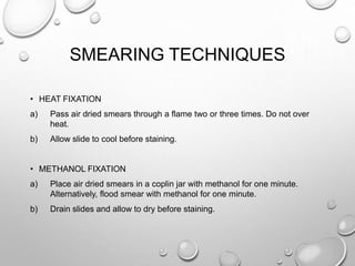
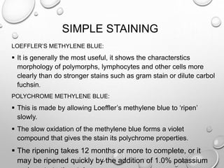
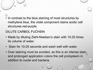
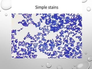
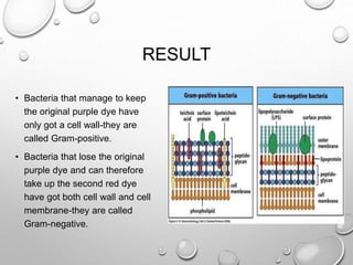
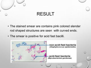
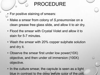
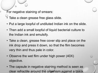
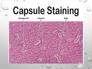
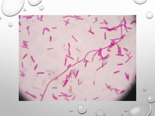
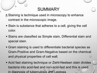
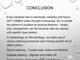
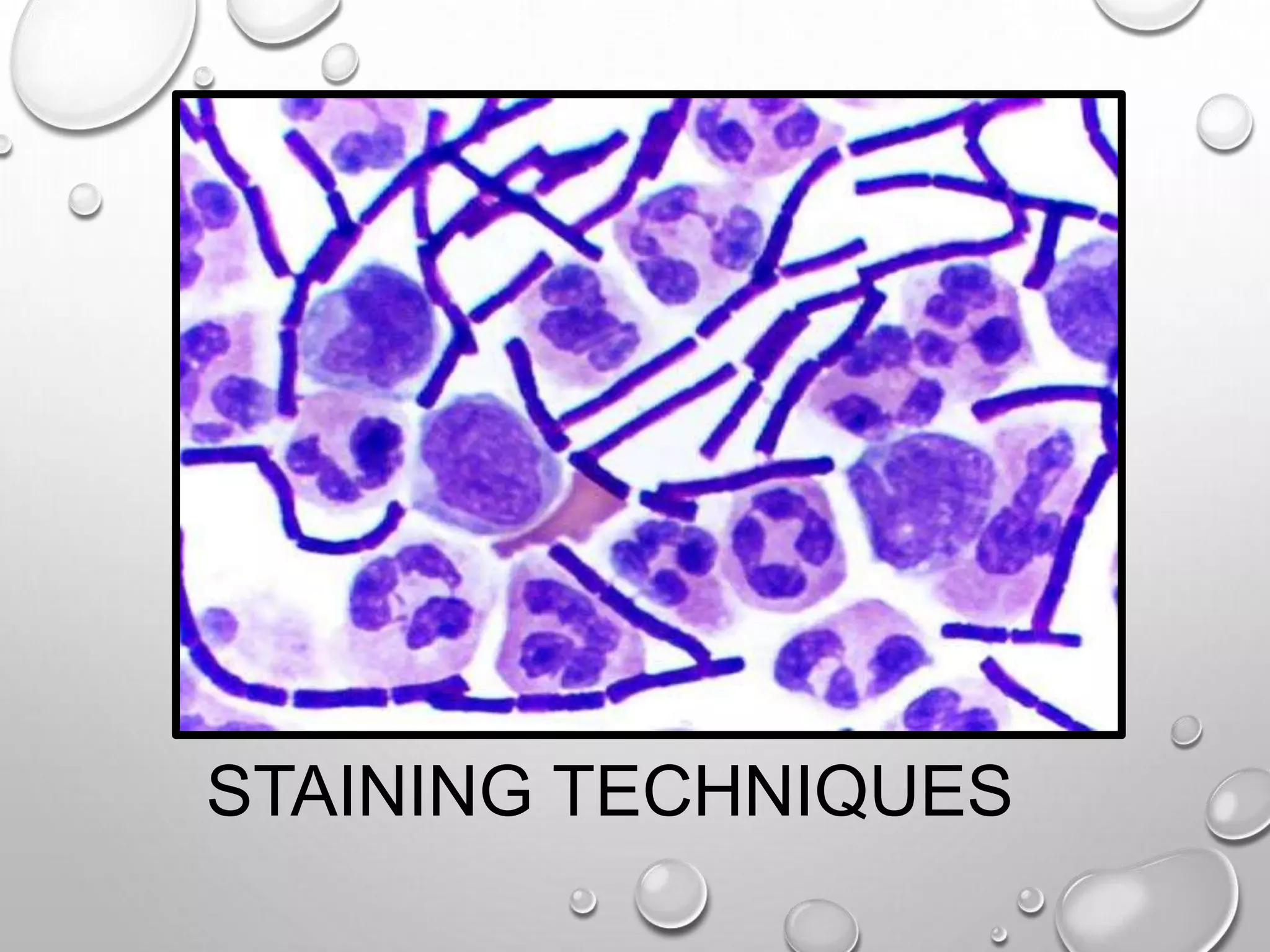
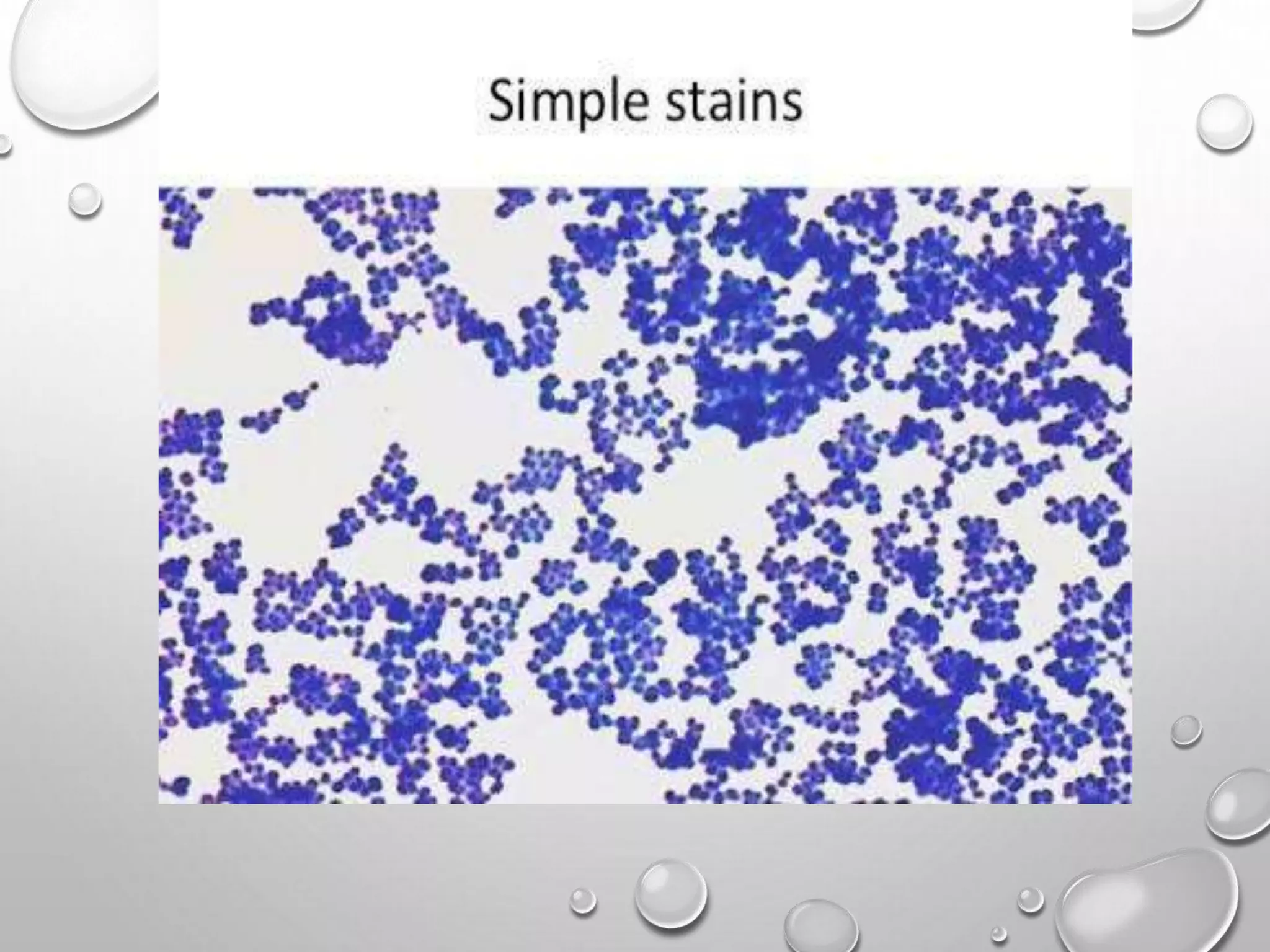
KEMBAR78
STAINING TECHNIQUES AND TYPES PROCEDURE. | PPTX
PADMASHREE INSTITUTE
OF
MANAGEMENT AND
SCIENCES
SUBMITTED BY:
SHYLESH MURTHY I A
M.SC BT, 1ST SEM
DEPT. OF BIOTECHNOLOGY, PIMS
TOPIC: STAINING TECHNIQUES
STAINING TECHNIQUES
INTRODUCTION
Staining is an auxiliary technique used in microscopy.
Stains and dyes are frequently used in biology and
medicine to highlight structure in biological tissues.
Stains may be used to define and examine bulk
tissues, cell populations or organelles with individual
cells.
Bacteria are microscopic organisms. They are mostly
colorless and to visualize them to study their structure,
shape and other structural characteristics.
TYPES OF STAINS
• ACIDIC
Negatively charged acid radicals imparts color in eosin, acid
fuchsine, malachite green, Indian ink.
• BASIC
Positively charged basic radicals combines with negatively
charged particles in cytoplasm and gives color.
e.g., Haematoxillin, Methylene blue, Crystal violet.
• NEURTAL
Both positively and negatively charged imparts different colors to
different components.
e.g., Geimsa’s stain and Leishman’s stain.
STAINING TECHNIQUES
POSITIVE STAINING
Where the actual cells are themselves colored and appear
in a clear background.
• Simple staining: a stain which provides color contrast but
gives same color to all bacteria an cells.
e.g., Malachite green and Crystal violet.
• Differential staining: a stain which imparts different colors
to different bacteria is called differential stain (which
contains more than one stain).
e.g., Gram’s stain, Acid fast staining and Endospore
staining.
• NEGATIVE STAINING :
Where the cells remain clear (uncolored) & the background is
colored to create a contrast to aid in the better visualization of the
image.
Nigrosin
Indian ink
BACTERIAL SMEAR PREPARATION
SMEAR : is a distribution of bacterial cells on a slide for the
purpose of viewing them under the microscope.
METHOD :
o Aseptically a small sample of the culture is spread over a
slide surface.
o This is then allowed to air dry.
o The next step is heat fixation to help the cells adhere to the
surface.
o The smear is now ready for staining.
SMEARING TECHNIQUES
• HEAT FIXATION
a) Pass air dried smears through a flame two or three times. Do not over
heat.
b) Allow slide to cool before staining.
• METHANOL FIXATION
a) Place air dried smears in a coplin jar with methanol for one minute.
Alternatively, flood smear with methanol for one minute.
b) Drain slides and allow to dry before staining.
SIMPLE STAINING
LOEFFLER’S METHYLENE BLUE:
 It is generally the most useful, it shows the characterstics
morphology of polymorphs, lymphocytes and other cells more
clearly than do stronger stains such as gram stain or dilute carbol
fuchsin.
POLYCHROME METHYLENE BLUE:
 This is made by allowing Loeffler’s methylene blue to ‘ripen’
slowly.
 The slow oxidation of the methylene blue forms a violet
compound that gives the stain its polychrome properties.
 The ripening takes 12 months or more to complete, or it
may be ripened quickly by the addition of 1.0% potassium
• In contrast to the blue staining of most structures by
methylene blue, the violet component stains acidic cell
structures red-purple.
DILUTE CARBOL FUCHSIN
• Made by diluting Ziehl-Neelsen’s stain with 10-20 times
its volume of water.
• Stain for 10-25 seconds and wash well with water.
• Over staining must be avoided, as this is an intense stain,
and prolonged application colors the cell protoplasm in
addition to nuclei and bacteria.
REQUIREMENTS
• Loefflers methylene blue
• Dilute Carbol Fuchsin
• Distilled water
• Compound microscope
• Cedar wood oil
• Fixed smear
PROCEDURE
• Make a thin smear on a slide.
• Heat fixes the smear by passing the slide 2-3 times gently over the
Bunsen flame with the smear side up.
• Pour Loeffler’s Methylene blue over the smear and allow it to stand
for 3 minutes.
• Wash the stained smear with water and air dry it.
• Observe the smear first under low power (10X) objective, and then
under oil immersion (100X) objective.
• Observe the presence of organisms and also the cellular content of
sample.
GRAM STAINING
• Gram staining is most widely used differential staining in
microbiology.
• It differentiates the bacteria into two groups:
Gram positive and Gram negative.
• Gram Positive Bacteria: They have a thick cell wall of
peptidoglycan and other polymers
• Peptidoglycan consists of interweaving filaments made up of
alternating N-acetylmuramic acid and N-acetylglucosamine
monomers.
• Gram Negative Bacteria: They have an outer membrane
of phospholipid and bacterial lipopolysaccharides outside
of their thin peptidoglycan layer.
• The outer membrane protects gram negative bacteria
againsts penicillin and lysozymes.
PROCEDURE OF GRAM STAINING
• It consists of four steps:
Primary staining: the smear is covered Crystal Violet, for
1 minute and washed with water.
Mordanting: it is then covered with Gram’s Iodine, kept for
1 minute, and washed with water.
Decolorization: the smear is covered with alcohol and is
washed with water immediately.
Counter staining: the smear is then covered with safarnin,
kept for 30 seconds and washed with water.
Observe under microscope.
RESULT
• Bacteria that manage to keep
the original purple dye have
only got a cell wall-they are
called Gram-positive.
• Bacteria that lose the original
purple dye and can therefore
take up the second red dye
have got both cell wall and cell
membrane-they are called
Gram-negative.
ACID-FAST STAINING
• The acid fast staining is a modification of Ehrlich’s (1882) method also
known as Ziehl-Neelsen stain.
• It is used for bacilli belonging to the genus Mycobacterium especially
Mycobacterium tuberculosis, Mycobacterium laprae and also for
Nocardia.
• Acid fastness of the acid-fast bacilli is attributed to the presence of
unsaponifiable wax fraction called mycolic acid in their cell wall.
• Basic requirements:
Primary and mordant staining with strong carbol fuchsin (red).
Decolorization with acid alcohol.
Counterstain with methylene blue.
PROCEDURE
• Make a smear. Air dry. Hear fix.
• Flood smear with Carbol-Fuchsin stain.
• Steam for 5 minutes. Add more of the stain as needed.
• Cool slide for 5 minutes. Wash with distilled water.
• Flood with acid alcohol (leave for 15 seconds).
• Tilt the slide 45⁰ over the sink and add acid alcohol drop wise
until the red color stops streaming from the smear.
• Rinse with distilled water.
• Add Loeffler’s methylene blue stain (Counter stain). Leave the
stain on smear for 15-20 seconds.
• Rinse the slide and let it dry.
• Use oil immersion objective to view.
RESULT
• The stained smear are contains pink colored slender
rod shaped structures are seen with curved ends.
• The smear is positive for acid fast bacilli.
CAPSULE STAINING
• The capsules serves as protective material by slowing
down or preventing penetration of chemicals and body
juices.
PRINCIPLE
• Chemically, the capsular material is a polysaccharide, a
glycoprotein or a polypeptide.
• Capsule staining is more difficult then other types of
differential staining procedures because he capsular
material are water soluble and may be dislodged and
removed with vigorous washing.
• The capsule is non-ionic, so that the dyes commonly
used will not bind to it. Two dyes, one acidic and one
PROCEDURE
• For positive staining of smears:
• Make a smear from colony of S.pneumoniae on a
clean grease free glass slide, and allow it to air dry.
• Flood the smear with Crystal Violet and allow it to
stain for 5-7 minutes.
• Wash the smear with 20% copper sulphate solution
and dry it.
• Observe the smear first under low power(10X)
objective, and then under oil immersion (100X)
objective.
• In the culture smear, the capsule is seen as a light
blue in contrast to the deep purple color of the cell.
For negative staining of smears:
• Take a clean grease free glass slide.
• Put a large loopful of undiluted Indian ink on the slide.
• Then add a small loopful of liquid bacterial culture to
the Indian ink and emulsify.
• Take a clean, grease free cover slip and place on the
ink drop and press it down, so that the flim becomes
very thin and thus pale in color.
• Observe the wet flim under high power (40X)
objective.
• The capsule in negative staining method is seen as
clear refractile around the organism against a black
ENDOSPORE STAINING
• Spores are highly resistant inactive forms.
• The morphology of bacterial endospores is best
observed in unstained wet flims under the phase
contrast microscope, where they appear as large ,
refractile, oval or spherical bodies within the bacterial
mother cells or else free from the bacteria.
• Different staining are available for staining of spores.
PROCEDURE
• Flims are dried and fixed with minimal flaming.
• Place the slide over a beaker of boiling water, resting it on the
rim with the bacterial flim uppermost.
• When, within several seconds, large droplets have condensed
on the under side of slide, flood it with 5% aqueous solution of
Malachite green and leave it for 1 minute while the water
continues to boil.
• Wash in cold water.
• Treat with 0.5% safranin and 0.05% basic fuchsin for 30
seconds.
• Wash and dry.
• This method colors the spores green and vegetative bacilli red.
SUMMARY
 Staining is technique used in microscopy to enhance
contrast in the microscopic image.
 Stain is substance that adheres to a cell, giving the cell
color.
 Stains are classified as Simple stain, Differential stain and
special stain.
 Gram staining is used to differentiate bacterial species as
Gram-Positive and Gram-Negative based on the chemical
and physical properties of cell wall.
 Acid fast staining technique or Ziehl-Neelsen stain divides
bacteria into acid-fast and non-acid-fast and this is used
in diagnosis of tuberculosis and Leprosy.
CONCLUSION
Every bacterial cell is individually colorless and hence
NOT VISIBLE under the light microscope. So, to enable
the person to visualize its physical features – shape,
size, arrangement, etc the bacterial cells are stained
with specific dyes (stains).
In bacteriology (or Microbiology), we make use of
various staining procedures each having specific set of
stains (dyes) –
Gram’s staining – Crystal violet, Iodine and Safranin
Capsule staining – Nigrosin and Indian ink
REFERNCE
• Prescott L.M, Harely T.P and Klein D.A, Microbiology,
7th edition P:30-52, Brown Publishers.
• www.biotechnika.com
• www.studymode.com

STAINING TECHNIQUES AND TYPES PROCEDURE.

  • 1.
    PADMASHREE INSTITUTE OF MANAGEMENT AND SCIENCES SUBMITTEDBY: SHYLESH MURTHY I A M.SC BT, 1ST SEM DEPT. OF BIOTECHNOLOGY, PIMS TOPIC: STAINING TECHNIQUES
  • 2.
  • 3.
    INTRODUCTION Staining is anauxiliary technique used in microscopy. Stains and dyes are frequently used in biology and medicine to highlight structure in biological tissues. Stains may be used to define and examine bulk tissues, cell populations or organelles with individual cells. Bacteria are microscopic organisms. They are mostly colorless and to visualize them to study their structure, shape and other structural characteristics.
  • 4.
    TYPES OF STAINS •ACIDIC Negatively charged acid radicals imparts color in eosin, acid fuchsine, malachite green, Indian ink. • BASIC Positively charged basic radicals combines with negatively charged particles in cytoplasm and gives color. e.g., Haematoxillin, Methylene blue, Crystal violet. • NEURTAL Both positively and negatively charged imparts different colors to different components. e.g., Geimsa’s stain and Leishman’s stain.
  • 5.
    STAINING TECHNIQUES POSITIVE STAINING Wherethe actual cells are themselves colored and appear in a clear background. • Simple staining: a stain which provides color contrast but gives same color to all bacteria an cells. e.g., Malachite green and Crystal violet. • Differential staining: a stain which imparts different colors to different bacteria is called differential stain (which contains more than one stain). e.g., Gram’s stain, Acid fast staining and Endospore staining.
  • 6.
    • NEGATIVE STAINING: Where the cells remain clear (uncolored) & the background is colored to create a contrast to aid in the better visualization of the image. Nigrosin Indian ink
  • 7.
    BACTERIAL SMEAR PREPARATION SMEAR: is a distribution of bacterial cells on a slide for the purpose of viewing them under the microscope. METHOD : o Aseptically a small sample of the culture is spread over a slide surface. o This is then allowed to air dry. o The next step is heat fixation to help the cells adhere to the surface. o The smear is now ready for staining.
  • 8.
    SMEARING TECHNIQUES • HEATFIXATION a) Pass air dried smears through a flame two or three times. Do not over heat. b) Allow slide to cool before staining. • METHANOL FIXATION a) Place air dried smears in a coplin jar with methanol for one minute. Alternatively, flood smear with methanol for one minute. b) Drain slides and allow to dry before staining.
  • 9.
    SIMPLE STAINING LOEFFLER’S METHYLENEBLUE:  It is generally the most useful, it shows the characterstics morphology of polymorphs, lymphocytes and other cells more clearly than do stronger stains such as gram stain or dilute carbol fuchsin. POLYCHROME METHYLENE BLUE:  This is made by allowing Loeffler’s methylene blue to ‘ripen’ slowly.  The slow oxidation of the methylene blue forms a violet compound that gives the stain its polychrome properties.  The ripening takes 12 months or more to complete, or it may be ripened quickly by the addition of 1.0% potassium
  • 10.
    • In contrastto the blue staining of most structures by methylene blue, the violet component stains acidic cell structures red-purple. DILUTE CARBOL FUCHSIN • Made by diluting Ziehl-Neelsen’s stain with 10-20 times its volume of water. • Stain for 10-25 seconds and wash well with water. • Over staining must be avoided, as this is an intense stain, and prolonged application colors the cell protoplasm in addition to nuclei and bacteria.
  • 11.
    REQUIREMENTS • Loefflers methyleneblue • Dilute Carbol Fuchsin • Distilled water • Compound microscope • Cedar wood oil • Fixed smear
  • 12.
    PROCEDURE • Make athin smear on a slide. • Heat fixes the smear by passing the slide 2-3 times gently over the Bunsen flame with the smear side up. • Pour Loeffler’s Methylene blue over the smear and allow it to stand for 3 minutes. • Wash the stained smear with water and air dry it. • Observe the smear first under low power (10X) objective, and then under oil immersion (100X) objective. • Observe the presence of organisms and also the cellular content of sample.
  • 14.
    GRAM STAINING • Gramstaining is most widely used differential staining in microbiology. • It differentiates the bacteria into two groups: Gram positive and Gram negative. • Gram Positive Bacteria: They have a thick cell wall of peptidoglycan and other polymers • Peptidoglycan consists of interweaving filaments made up of alternating N-acetylmuramic acid and N-acetylglucosamine monomers.
  • 15.
    • Gram NegativeBacteria: They have an outer membrane of phospholipid and bacterial lipopolysaccharides outside of their thin peptidoglycan layer. • The outer membrane protects gram negative bacteria againsts penicillin and lysozymes.
  • 16.
    PROCEDURE OF GRAMSTAINING • It consists of four steps: Primary staining: the smear is covered Crystal Violet, for 1 minute and washed with water. Mordanting: it is then covered with Gram’s Iodine, kept for 1 minute, and washed with water. Decolorization: the smear is covered with alcohol and is washed with water immediately. Counter staining: the smear is then covered with safarnin, kept for 30 seconds and washed with water. Observe under microscope.
  • 17.
    RESULT • Bacteria thatmanage to keep the original purple dye have only got a cell wall-they are called Gram-positive. • Bacteria that lose the original purple dye and can therefore take up the second red dye have got both cell wall and cell membrane-they are called Gram-negative.
  • 18.
    ACID-FAST STAINING • Theacid fast staining is a modification of Ehrlich’s (1882) method also known as Ziehl-Neelsen stain. • It is used for bacilli belonging to the genus Mycobacterium especially Mycobacterium tuberculosis, Mycobacterium laprae and also for Nocardia. • Acid fastness of the acid-fast bacilli is attributed to the presence of unsaponifiable wax fraction called mycolic acid in their cell wall. • Basic requirements: Primary and mordant staining with strong carbol fuchsin (red). Decolorization with acid alcohol. Counterstain with methylene blue.
  • 19.
    PROCEDURE • Make asmear. Air dry. Hear fix. • Flood smear with Carbol-Fuchsin stain. • Steam for 5 minutes. Add more of the stain as needed. • Cool slide for 5 minutes. Wash with distilled water. • Flood with acid alcohol (leave for 15 seconds). • Tilt the slide 45⁰ over the sink and add acid alcohol drop wise until the red color stops streaming from the smear. • Rinse with distilled water. • Add Loeffler’s methylene blue stain (Counter stain). Leave the stain on smear for 15-20 seconds. • Rinse the slide and let it dry. • Use oil immersion objective to view.
  • 20.
    RESULT • The stainedsmear are contains pink colored slender rod shaped structures are seen with curved ends. • The smear is positive for acid fast bacilli.
  • 21.
    CAPSULE STAINING • Thecapsules serves as protective material by slowing down or preventing penetration of chemicals and body juices. PRINCIPLE • Chemically, the capsular material is a polysaccharide, a glycoprotein or a polypeptide. • Capsule staining is more difficult then other types of differential staining procedures because he capsular material are water soluble and may be dislodged and removed with vigorous washing. • The capsule is non-ionic, so that the dyes commonly used will not bind to it. Two dyes, one acidic and one
  • 22.
    PROCEDURE • For positivestaining of smears: • Make a smear from colony of S.pneumoniae on a clean grease free glass slide, and allow it to air dry. • Flood the smear with Crystal Violet and allow it to stain for 5-7 minutes. • Wash the smear with 20% copper sulphate solution and dry it. • Observe the smear first under low power(10X) objective, and then under oil immersion (100X) objective. • In the culture smear, the capsule is seen as a light blue in contrast to the deep purple color of the cell.
  • 23.
    For negative stainingof smears: • Take a clean grease free glass slide. • Put a large loopful of undiluted Indian ink on the slide. • Then add a small loopful of liquid bacterial culture to the Indian ink and emulsify. • Take a clean, grease free cover slip and place on the ink drop and press it down, so that the flim becomes very thin and thus pale in color. • Observe the wet flim under high power (40X) objective. • The capsule in negative staining method is seen as clear refractile around the organism against a black
  • 25.
    ENDOSPORE STAINING • Sporesare highly resistant inactive forms. • The morphology of bacterial endospores is best observed in unstained wet flims under the phase contrast microscope, where they appear as large , refractile, oval or spherical bodies within the bacterial mother cells or else free from the bacteria. • Different staining are available for staining of spores.
  • 26.
    PROCEDURE • Flims aredried and fixed with minimal flaming. • Place the slide over a beaker of boiling water, resting it on the rim with the bacterial flim uppermost. • When, within several seconds, large droplets have condensed on the under side of slide, flood it with 5% aqueous solution of Malachite green and leave it for 1 minute while the water continues to boil. • Wash in cold water. • Treat with 0.5% safranin and 0.05% basic fuchsin for 30 seconds. • Wash and dry. • This method colors the spores green and vegetative bacilli red.
  • 28.
    SUMMARY  Staining istechnique used in microscopy to enhance contrast in the microscopic image.  Stain is substance that adheres to a cell, giving the cell color.  Stains are classified as Simple stain, Differential stain and special stain.  Gram staining is used to differentiate bacterial species as Gram-Positive and Gram-Negative based on the chemical and physical properties of cell wall.  Acid fast staining technique or Ziehl-Neelsen stain divides bacteria into acid-fast and non-acid-fast and this is used in diagnosis of tuberculosis and Leprosy.
  • 29.
    CONCLUSION Every bacterial cellis individually colorless and hence NOT VISIBLE under the light microscope. So, to enable the person to visualize its physical features – shape, size, arrangement, etc the bacterial cells are stained with specific dyes (stains). In bacteriology (or Microbiology), we make use of various staining procedures each having specific set of stains (dyes) – Gram’s staining – Crystal violet, Iodine and Safranin Capsule staining – Nigrosin and Indian ink
  • 30.
    REFERNCE • Prescott L.M,Harely T.P and Klein D.A, Microbiology, 7th edition P:30-52, Brown Publishers. • www.biotechnika.com • www.studymode.com