Download to read offline


![International Research Journal of Engineering and Technology (IRJET) e-ISSN: 2395-0056
Volume: 11 Issue: 02 | Feb 2024 www.irjet.net p-ISSN: 2395-0072
© 2024, IRJET | Impact Factor value: 8.226 | ISO 9001:2008 Certified Journal | Page 79
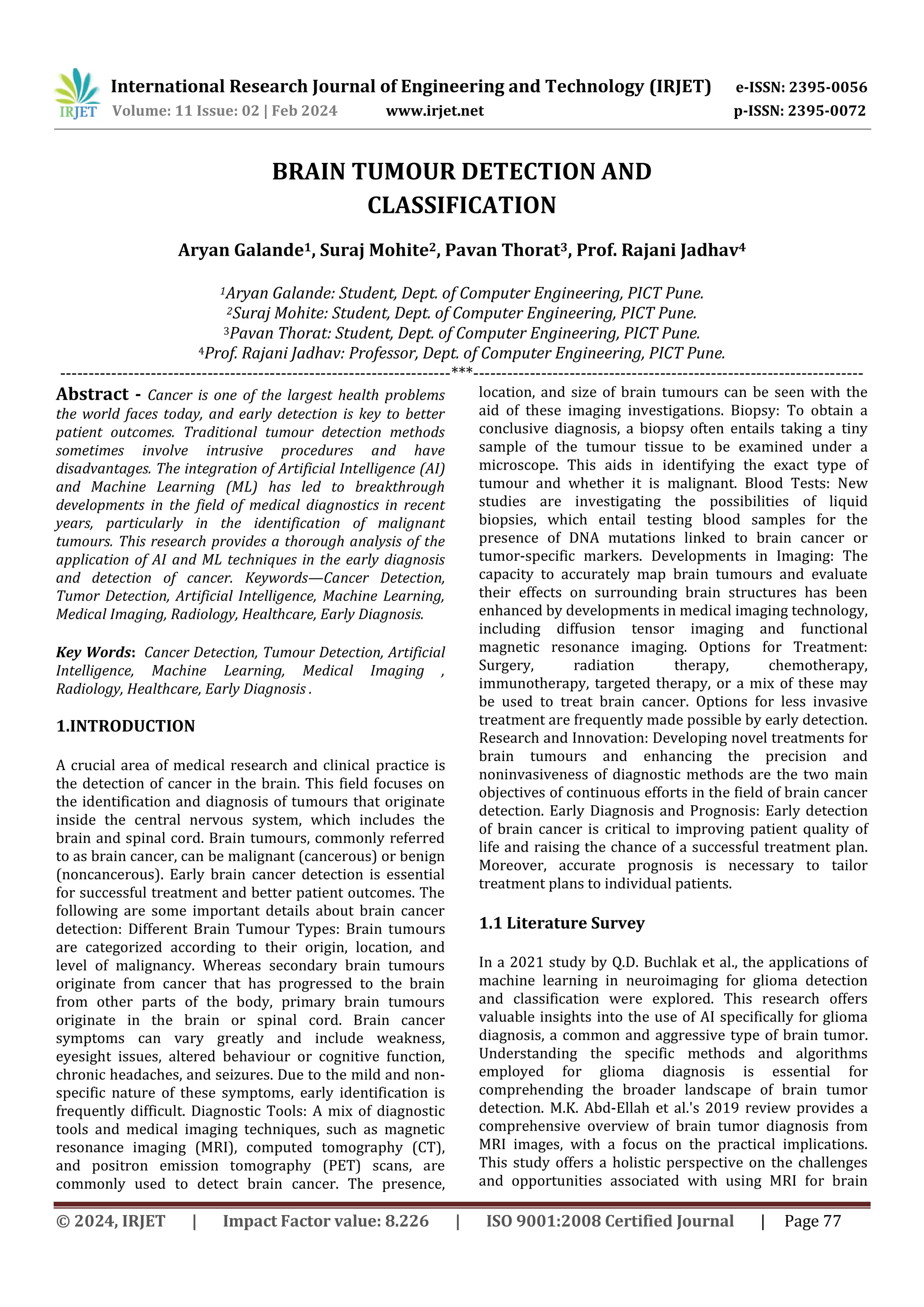
The chosen dataset includes two types of data:-
1)Training Data
2)Testing Data
Each of these divisions of datasets includes MRI Scansof
Human Brain having no tumor and MRI Scans of Human
Brain having Pituitary Tumor.
The SVM Model is trained on the training data of chosen
dataset, Fig 2 shows results of testing of SVM model on the
testing data of chosen dataset for no tumor.
Fig. 3. Samples tested for pituitary tumor.
Fig 3 shows results of testing of SVM model on the
testingdata of chosen dataset for pituitary tumor.
Fig. 4. Testing Results.
After testing the model, we got the training score of
0.9887 andtesting score of 0.9592.
3. CONCLUSIONS
In conclusion, a promising direction in the field of medical
diagnostics is the application of Support Vector Machines
(SVM) for brain tumour detection. SVMs are a useful tool
for accurate classification, but there are issues that need to
be resolved, including issues with generalization, data
limitations, and ethics.
Investigating deep learning methods, multimodal data
fusion, and real-time detection systems, among other
possibilities, can significantly improve the efficiency and
[1] Q.D. Buchlak et al. Machine learning applications to
neuroimaging for glioma detection and classification: an
artificial intelligence augmented systematic review J. Clin.
Neurosci. (2021)
[2] M.K. Abd-Ellah et al. A review on brain tumor diagnosis
from MRI images: practical implications, key
achievements, and lessons learned Magn. Reson. Imag.
(2019)
[3] M.K. Abd-Ellah et al. A review on brain tumor diagnosis
from MRI images: practical implications, key
achievements, and lessons learned Magn. Reson. Imag.
(2019)
[4] V.P. Grover et al. Magnetic resonance imaging:
principles and techniques: lessons for clinicians Journal of
clinical and experimental hepatology (2015)
[5]. H. Tang et al. MRI brain image segmentation by
multiresolution edge detection and region selection
Comput. Med. Imag. Graph. (2000)
[6] K. Somasundaram et al. Fully automatic brain
extraction algorithm for axial T2-weighted magnetic
resonance images Comput. Biol. Med. (2010)
accuracy of tumour detection. Moreover, collaboration
between AI and medical experts is necessary for the
successful integration of SVM-based tumour detection in
clinical settings. These developments herald a bright
future for improving brain tumour identification, which
will eventually lead to better patient outcomes and care.
REFERENCES](https://image.slidesharecdn.com/irjet-v11i214-241030175856-bf7b2762/85/BRAIN-TUMOUR-DETECTION-AND-CLASSIFICATION-3-320.jpg)
![International Research Journal of Engineering and Technology (IRJET) e-ISSN: 2395-0056
Volume: 11 Issue: 02 | Feb 2024 www.irjet.net p-ISSN: 2395-0072
© 2024, IRJET | Impact Factor value: 8.226 | ISO 9001:2008 Certified Journal | Page 79
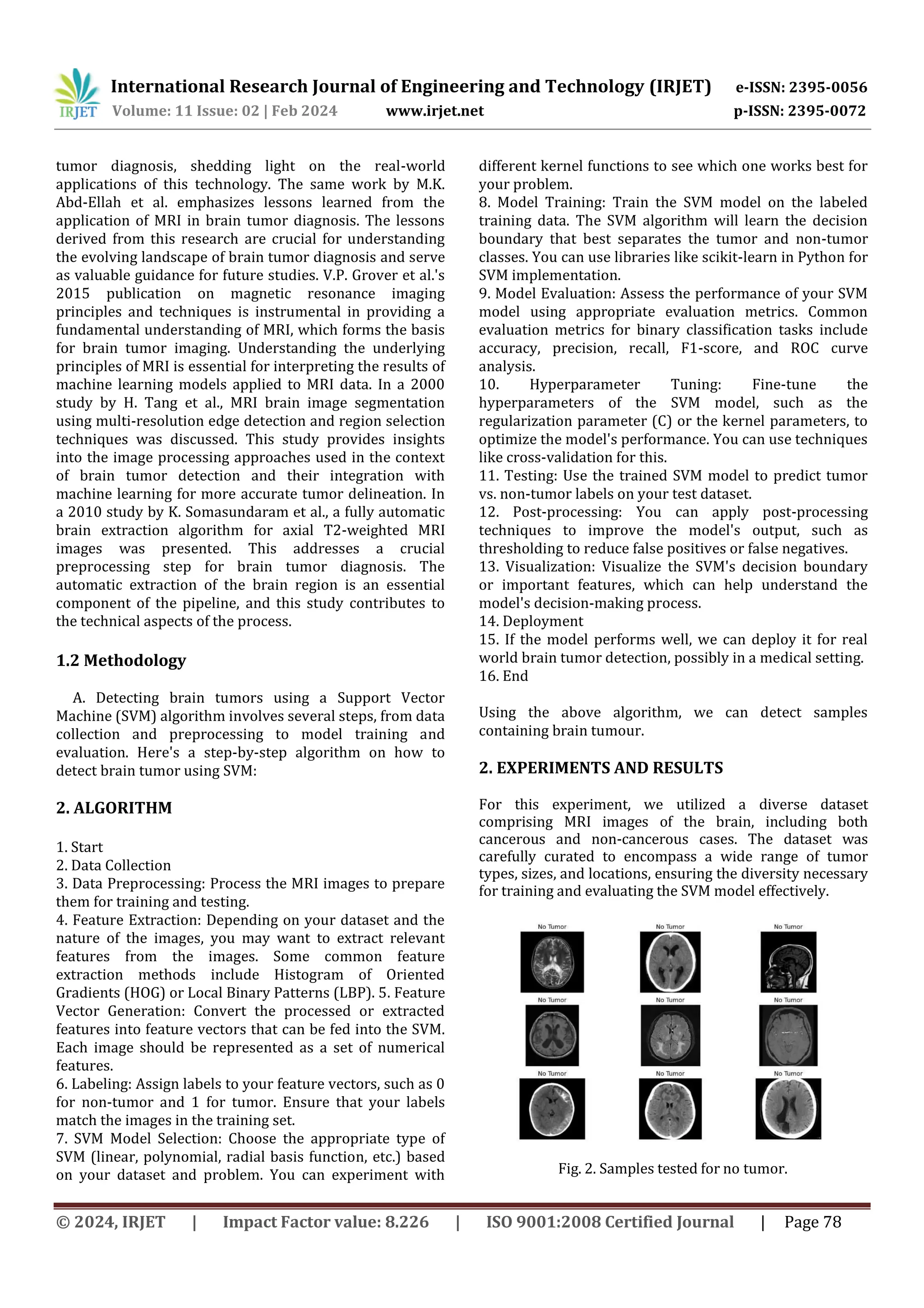
The chosen dataset includes two types of data:-
1)Training Data
2)Testing Data
Each of these divisions of datasets includes MRI Scansof
Human Brain having no tumor and MRI Scans of Human
Brain having Pituitary Tumor.
The SVM Model is trained on the training data of chosen
dataset, Fig 2 shows results of testing of SVM model on the
testing data of chosen dataset for no tumor.
Fig. 3. Samples tested for pituitary tumor.
Fig 3 shows results of testing of SVM model on the
testingdata of chosen dataset for pituitary tumor.
Fig. 4. Testing Results.
After testing the model, we got the training score of
0.9887 andtesting score of 0.9592.
3. CONCLUSIONS
In conclusion, a promising direction in the field of medical
diagnostics is the application of Support Vector Machines
(SVM) for brain tumour detection. SVMs are a useful tool
for accurate classification, but there are issues that need to
be resolved, including issues with generalization, data
limitations, and ethics.
Investigating deep learning methods, multimodal data
fusion, and real-time detection systems, among other
possibilities, can significantly improve the efficiency and
[1] Q.D. Buchlak et al. Machine learning applications to
neuroimaging for glioma detection and classification: an
artificial intelligence augmented systematic review J. Clin.
Neurosci. (2021)
[2] M.K. Abd-Ellah et al. A review on brain tumor diagnosis
from MRI images: practical implications, key
achievements, and lessons learned Magn. Reson. Imag.
(2019)
[3] M.K. Abd-Ellah et al. A review on brain tumor diagnosis
from MRI images: practical implications, key
achievements, and lessons learned Magn. Reson. Imag.
(2019)
[4] V.P. Grover et al. Magnetic resonance imaging:
principles and techniques: lessons for clinicians Journal of
clinical and experimental hepatology (2015)
[5]. H. Tang et al. MRI brain image segmentation by
multiresolution edge detection and region selection
Comput. Med. Imag. Graph. (2000)
[6] K. Somasundaram et al. Fully automatic brain
extraction algorithm for axial T2-weighted magnetic
resonance images Comput. Biol. Med. (2010)
accuracy of tumour detection. Moreover, collaboration
between AI and medical experts is necessary for the
successful integration of SVM-based tumour detection in
clinical settings. These developments herald a bright
future for improving brain tumour identification, which
will eventually lead to better patient outcomes and care.
REFERENCES](https://image.slidesharecdn.com/irjet-v11i214-241030175856-bf7b2762/75/BRAIN-TUMOUR-DETECTION-AND-CLASSIFICATION-3-2048.jpg)

This document presents a study on the application of artificial intelligence (AI) and machine learning (ML), specifically using support vector machines (SVM), for the early detection and classification of brain tumors. It emphasizes the importance of early detection in improving treatment outcomes and reviews existing methodologies and technologies for brain tumor diagnosis. The findings indicate high accuracy in tumor detection, with the SVM model achieving a training score of 0.9887 and a testing score of 0.9592.


