Download to read offline
![International Research Journal of Engineering and Technology (IRJET) e-ISSN: 2395-0056
Volume: 11 Issue: 02 | Feb 2024 www.irjet.net p-ISSN: 2395-0072
© 2024, IRJET | Impact Factor value: 8.226 | ISO 9001:2008 Certified Journal | Page 80
Breast Cancer Detection using Computer Vision
Janhavi Tingre
School of Computer
Engineering & Technology
MIT World Peace University,
Pune, Maharashtra
Vaishnavi Mundada
School of Computer
Engineering & Technology
MIT World Peace University,
Pune, Maharashtra
Harsh Shelke
School of Computer
Engineering & Technology
MIT World Peace University,
Pune, Maharashtra
Ayush Chaudhary
School of Computer
Engineering & Technology
MIT World Peace University,
Pune, Maharashtra
--------------------------------------------------------------------***-----------------------------------------------------------------
Abstract— Breast cancer is among the main reasons
why women die worldwide. Breast cancer mortality
rates and treatment expenses can be decreased with
early detection and diagnosis. In this effort, we have
put forth a novel, affordable, computer vision-based
method for detecting and diagnosing breast cancer.
Convolutional neural networks are also used for
medical image classification. The proposed model is
a very simple and cost effective approach with high
accuracy and useful outcomes. We have also
explored the different challenges faced and the
future scope of the project.
Keywords—Breast Cancer, Computer vision,
Convolutional neural network, Detection
I. INTRODUCTION
Overtaking lung cancer, breast cancer is the most
common cancer among women. In India the survival rate
for breast cancer patients is about 60% as compared to
90% in the United States, for the last five years [1]. By
enhancing treatment options, early detection methods,
awareness campaigns, and better diagnostics, we can
increase these survival rates.
Because of its simplicity and practicability, ultrasound
has become a standard tool for diagnosing breast
disorders. The findings of B-mode ultrasonography, on
the other hand, are related to the level of expertise of
doctors, poor image quality, benign presentations of
malignant tumors, and visual fatigue or neglect on the
part of observers [2]. If a huge number of ultrasound
mammary images are manually examined, there will be
significant flaws. Misdiagnosis is common when lesions
that should be properly diagnosed are missed by
radiologists [2].
CALC (calcification), CIRC (circumcised masses), SPIC
(speculated masses), MISC (other ill-defined masses),
ARCH (architectural distortion), and ASYM (asymmetry)
are the six types of breast cancer [3]. In this paper,
Invasive Ductal Carcinoma (IDC), one of the most
prevalent forms of breast cancer, is one that we are
finding.
There is improvement in the field of diagnosis due to
evolving technology. Convolutional neural network is the
most widely used machine learning algorithm in the field
of medical image analysis [4]. The fundamental reason
for this is because CNN exactly fits the two-dimensional
structure of the image in structure and uses this spatial
relationship as the algorithm's direct input value [4].
II. OBJECTIVES
The main objective of this project is to design a
computer vision system that can help with early
detection of breast cancer. In this project, we have
explored computer vision as an image preprocessing
technique. Along with it, convolutional neural networks
are used for image classification. System architecture is
shown below fig.1
Fig.1 System Architecture](https://image.slidesharecdn.com/irjet-v11i215-241030163405-1512c3b6/85/Breast-Cancer-Detection-using-Computer-Vision-1-320.jpg)
![© 2024, IRJET | Impact Factor value: 8.226 | ISO 9001:2008 Certified Journal | Page 81
III. LITERATURE SURVEY
For the literature survey, we studied multiple research
papers based on breast cancer detection and different
methods to achieve it. The descriptive details about the
same have been mentioned in the sections below.
A. This research paper presents a method for breast
cancer (BC) identification and categorization using K-
nearest neighbor (KNN), classification of
thermographic images using (SVM) with (SMO), and
detection of BC nuclei using Stacked Sparse
Autoencoder (SSAE). The study suggests that training
on additional datasets can improve the accuracy of
the model [5]. Furthermore, the authors emphasize
the importance of verifying medical photos using a
broad range of applications since some tumor
diagnoses can be challenging [5]. Overall, the
proposed approach can aid in the accurate detection
and diagnosis of breast cancer.
B. In this research paper, they used different machine
learning algorithms, including multilayer perceptron
neural networks (MLPNNs), (SVMs), and (LDA), in
various applications of pattern recognition. The study
highlights a major challenge in this field, which is the
unbalanced dataset problem, where one class has
significantly more samples than another [6]. The
authors explore different strategies to address this
issue, such as resampling techniques and adjusting
the cost matrix [6]. Overall, the findings of this
research provide insights into the selection of
appropriate algorithms and techniques to improve
the accuracy of pattern recognition in real-world
scenarios with unbalanced datasets.
C. This research paper discusses the Breast Imaging
Report and Data System (BI-RADS) as a standardized
tool for mammography reporting and interpretation.
The authors highlight the various categories within
the BI-RADS system, including BI-RADS 0, 1, and 2,
where BI-RADS 2 denotes a benign finding with a
probability of malignancy of 0% [7]. The study
emphasizes the importance of considering the
possibility of false-negative or false-positive results
in interpreting mammography findings and
recommends that radiologists incorporate this into
their decision-making process. The authors also
explore the role of BI-RADS in facilitating
communication between radiologists and referring
physicians and guiding patient management [7].
Overall, the findings of this research contribute to the
understanding and implementation of the BI-RADS
system in clinical practice, improving breast cancer
detection and diagnosis.
D. This research paper presents the use of pre-trained
deep neural network (DNN) models, including
ResNet, Inception-V3Net, and ShuffleNet, for
conducting binary and multi-class classifications. The
study highlights the importance of labeled data for
training these models effectively. However, the
authors also acknowledge that the lack of
interpretability in DNN models can make it
challenging to identify false positives or false
negatives [8]. The research demonstrates the
potential of pre-trained DNNs in achieving high
accuracy in classification tasks, particularly in image
analysis. The findings of this study contribute to the
understanding of the practical applications of pre-
trained DNNs and the challenges associated with
their use in real-world scenarios [8].
E. In this research paper, system evaluates computing
approaches for breast cancer detection based on CAD
using mammogram images, focusing on threshold-
based and region-based segmentation. However, the
increased computational challenges associated with
machine learning (ML) classifiers based on deep
learning (DL) as the number of layers increases have
been identified as a research gap [9]. DL-based
classifiers have shown great potential in breast
cancer detection, but their computational challenges
make them less practical for clinical settings [9].
Thus, more research is needed to develop efficient
and robust computing approaches for breast cancer
detection using mammogram images.
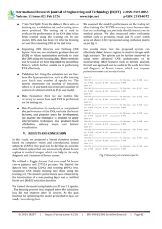
IV. METHODOLOGY
Breast Cancer is an ordinary form of cancer among
women and immediate assessment is important for
successful cure. Convolutional Neural Networks have
shown encouraging results in the prognosis of breast
cancer using Whole Source Images (WSI). Below is the
normal methodology of detection of Breast Cancer:
● Assembling the Data: The dataset should have a
gargantuan variety of Whole Source Images
ranging from malignant to innocuous tumor
images. Consequently, we prepare and construct
tiles of those Whole Source Images.
● Data Segregation: Further, we separate the
aforementioned tiles into cancerous and non-
cancerous arrays respectively.
● Data Pre-Processing: Here we preprocess the
data using the open computer vision
library(cv2). The open computer vision library
(cv2) is primarily concerned with image
processing, video recording, and analysis, which
includes functions like face and object detection.
Here, we make use of this library to resize and
redimension all of the photos. Combination of
the arrays: We conjoin the two above mentioned
arrays into a single array.
International Research Journal of Engineering and Technology (IRJET) e-ISSN: 2395-0056
Volume: 11 Issue: 02 | Feb 2024 www.irjet.net p-ISSN: 2395-0072](https://image.slidesharecdn.com/irjet-v11i215-241030163405-1512c3b6/85/Breast-Cancer-Detection-using-Computer-Vision-2-320.jpg)
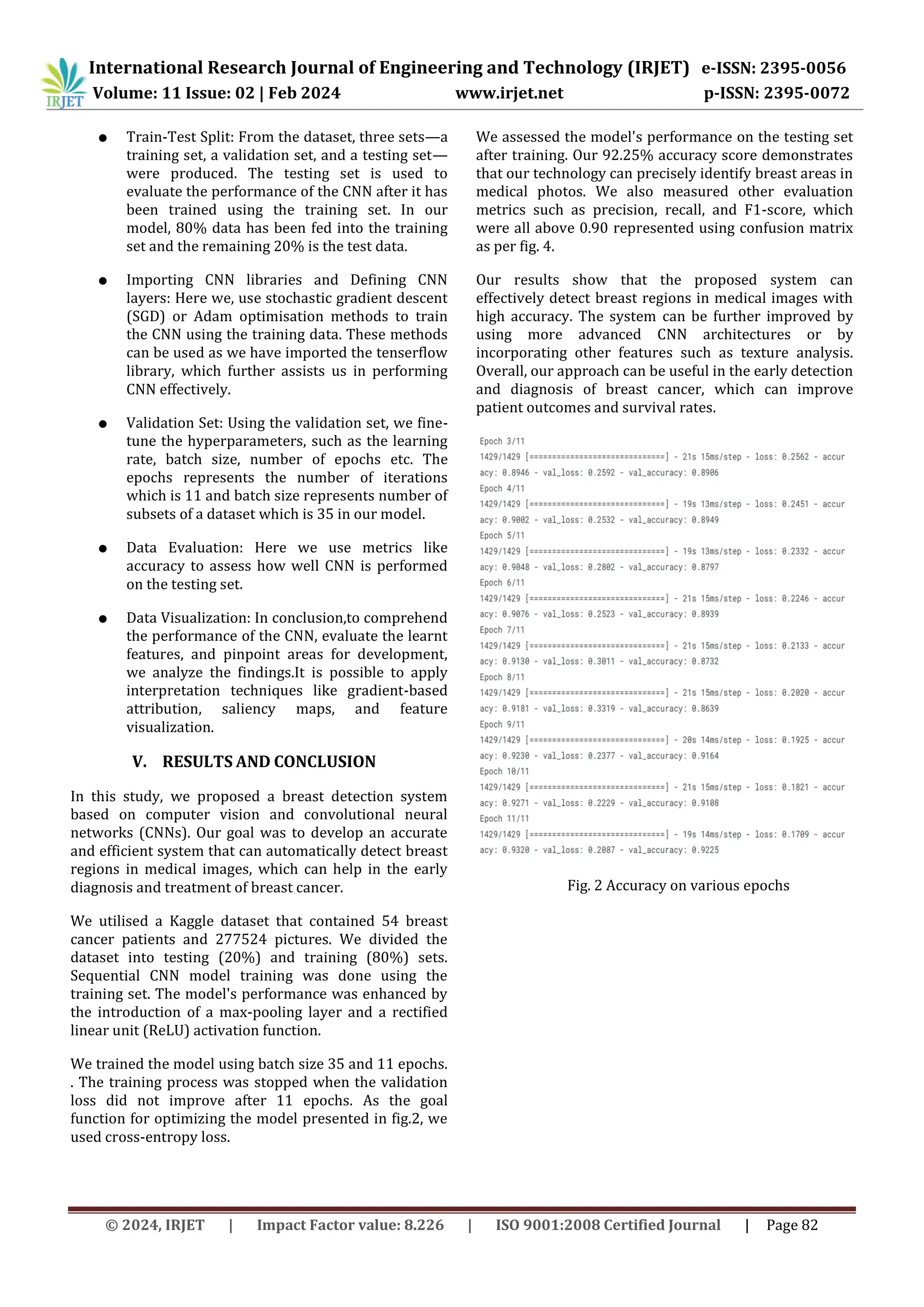
![© 2024, IRJET | Impact Factor value: 8.226 | ISO 9001:2008 Certified Journal | Page 83
Fig. 3. Performance of proposed model
Fig. 4. Confusion matrix
REFERENCES
[1] R. Guzman-Cabrera, J.R. Guzm ´ an-Sep ´ ulveda, M. ´
313 Torres-Cisneros, D.A. May-Arrioja, J. Ruiz-
Pinales, 314 O.G. Ibarra-Manzano, et al., Digital
image processing 315 technique for breast cancer
detection,.
[2] International 316 Journal of Thermophysics 34
(2013), 1519–1531.
[3] Jackson V. P., Hendrick R. E., Feig S. A., et al. Imaging
of the radiographically dense breast.[J]. Radiology,
1993, 188(2):297-301.
[4] Sethy, P. K., Pandey, C., Khan, D. M. R., Behera, S. K.,
Vijaykumar, K., & Panigrahi, D. S. (2021). A cost-
effective computer-vision based breast cancer
diagnosis. Journal of Intelligent & Fuzzy Systems, 1–
11. doi:10.3233/jifs-189848
[5] Yunchao, G., & Jiayao, Y. (2019). Application of
Computer Vision and Deep Learning in Breast
Cancer Assisted Diagnosis. Proceedings of the 3rd
International Conference on Machine Learning and
Soft Computing - ICMLC 2019.
doi:10.1145/3310986.3311010
[6] Sethy, Prabira Kumar et al. ‘A Cost-effective
Computer-vision Based Breast Cancer Diagnosis’. 1
Jan. 2021 : 5253 – 5263.
[7] Jalalian A, Mashohor S, Mahmud R, Karasfi B, Saripan
MIB, Ramli ARB. Foundation and methodologies in
computer-aided diagnosis systems for breast cancer
detection. EXCLI J. 2017;16:113-137. Published
2017 Feb 20. doi:10.17179/excli2016-701
[8] Al-Tam, Riyadh & Narangale, Sachin. (2021). Breast
Cancer Detection and Diagnosis Using Machine
Learning: A Survey. JOURNAL OF SCIENTIFIC
RESEARCH. 65. 265-285.
10.37398/JSR.2021.650532.
[9] Al-Tam, Riyadh & Narangale, Sachin. (2021). Breast
Cancer Detection and Diagnosis Using Machine
Learning: A Survey. JOURNAL OF SCIENTIFIC
RESEARCH. 65. 265-285.
10.37398/JSR.2021.650532.
[10] Aljuaid H, Alturki N, Alsubaie N, Cavallaro L, Liotta A.
Computer-aided diagnosis for breast cancer
classification using deep neural networks and
transfer learning. Comput Methods Programs
Biomed. 2022;223:106951.
doi:10.1016/j.cmpb.2022.106951
[11] Asma Zizaan, Ali Idri. (2023) Machine learning
based Breast Cancer screening: trends, challenges,
and opportunities. Computer Methods in
Biomechanics and Biomedical Engineering: Imaging
& Visualization 0:0, pages 1-21.
[12] Pawar, R., Ghumbre, S., & Deshmukh, R. (2019).
Visual Similarity Using Convolution Neural Network
over Textual Similarity in Content- Based
Recommender System. International Journal of
Advanced Science and Technology, 27, 137 - 147.
[13] Pawar, R., Ghumbre, S., & Deshmukh, R. (2020). A
Hybrid Approach towards Improving Performance
International Research Journal of Engineering and Technology (IRJET) e-ISSN: 2395-0056
Volume: 11 Issue: 02 | Feb 2024 www.irjet.net p-ISSN: 2395-0072](https://image.slidesharecdn.com/irjet-v11i215-241030163405-1512c3b6/85/Breast-Cancer-Detection-using-Computer-Vision-4-320.jpg)

![International Research Journal of Engineering and Technology (IRJET) e-ISSN: 2395-0056
Volume: 11 Issue: 02 | Feb 2024 www.irjet.net p-ISSN: 2395-0072
© 2024, IRJET | Impact Factor value: 8.226 | ISO 9001:2008 Certified Journal | Page 80
Breast Cancer Detection using Computer Vision
Janhavi Tingre
School of Computer
Engineering & Technology
MIT World Peace University,
Pune, Maharashtra
Vaishnavi Mundada
School of Computer
Engineering & Technology
MIT World Peace University,
Pune, Maharashtra
Harsh Shelke
School of Computer
Engineering & Technology
MIT World Peace University,
Pune, Maharashtra
Ayush Chaudhary
School of Computer
Engineering & Technology
MIT World Peace University,
Pune, Maharashtra
--------------------------------------------------------------------***-----------------------------------------------------------------
Abstract— Breast cancer is among the main reasons
why women die worldwide. Breast cancer mortality
rates and treatment expenses can be decreased with
early detection and diagnosis. In this effort, we have
put forth a novel, affordable, computer vision-based
method for detecting and diagnosing breast cancer.
Convolutional neural networks are also used for
medical image classification. The proposed model is
a very simple and cost effective approach with high
accuracy and useful outcomes. We have also
explored the different challenges faced and the
future scope of the project.
Keywords—Breast Cancer, Computer vision,
Convolutional neural network, Detection
I. INTRODUCTION
Overtaking lung cancer, breast cancer is the most
common cancer among women. In India the survival rate
for breast cancer patients is about 60% as compared to
90% in the United States, for the last five years [1]. By
enhancing treatment options, early detection methods,
awareness campaigns, and better diagnostics, we can
increase these survival rates.
Because of its simplicity and practicability, ultrasound
has become a standard tool for diagnosing breast
disorders. The findings of B-mode ultrasonography, on
the other hand, are related to the level of expertise of
doctors, poor image quality, benign presentations of
malignant tumors, and visual fatigue or neglect on the
part of observers [2]. If a huge number of ultrasound
mammary images are manually examined, there will be
significant flaws. Misdiagnosis is common when lesions
that should be properly diagnosed are missed by
radiologists [2].
CALC (calcification), CIRC (circumcised masses), SPIC
(speculated masses), MISC (other ill-defined masses),
ARCH (architectural distortion), and ASYM (asymmetry)
are the six types of breast cancer [3]. In this paper,
Invasive Ductal Carcinoma (IDC), one of the most
prevalent forms of breast cancer, is one that we are
finding.
There is improvement in the field of diagnosis due to
evolving technology. Convolutional neural network is the
most widely used machine learning algorithm in the field
of medical image analysis [4]. The fundamental reason
for this is because CNN exactly fits the two-dimensional
structure of the image in structure and uses this spatial
relationship as the algorithm's direct input value [4].
II. OBJECTIVES
The main objective of this project is to design a
computer vision system that can help with early
detection of breast cancer. In this project, we have
explored computer vision as an image preprocessing
technique. Along with it, convolutional neural networks
are used for image classification. System architecture is
shown below fig.1
Fig.1 System Architecture](https://image.slidesharecdn.com/irjet-v11i215-241030163405-1512c3b6/75/Breast-Cancer-Detection-using-Computer-Vision-1-2048.jpg)
![© 2024, IRJET | Impact Factor value: 8.226 | ISO 9001:2008 Certified Journal | Page 81
III. LITERATURE SURVEY
For the literature survey, we studied multiple research
papers based on breast cancer detection and different
methods to achieve it. The descriptive details about the
same have been mentioned in the sections below.
A. This research paper presents a method for breast
cancer (BC) identification and categorization using K-
nearest neighbor (KNN), classification of
thermographic images using (SVM) with (SMO), and
detection of BC nuclei using Stacked Sparse
Autoencoder (SSAE). The study suggests that training
on additional datasets can improve the accuracy of
the model [5]. Furthermore, the authors emphasize
the importance of verifying medical photos using a
broad range of applications since some tumor
diagnoses can be challenging [5]. Overall, the
proposed approach can aid in the accurate detection
and diagnosis of breast cancer.
B. In this research paper, they used different machine
learning algorithms, including multilayer perceptron
neural networks (MLPNNs), (SVMs), and (LDA), in
various applications of pattern recognition. The study
highlights a major challenge in this field, which is the
unbalanced dataset problem, where one class has
significantly more samples than another [6]. The
authors explore different strategies to address this
issue, such as resampling techniques and adjusting
the cost matrix [6]. Overall, the findings of this
research provide insights into the selection of
appropriate algorithms and techniques to improve
the accuracy of pattern recognition in real-world
scenarios with unbalanced datasets.
C. This research paper discusses the Breast Imaging
Report and Data System (BI-RADS) as a standardized
tool for mammography reporting and interpretation.
The authors highlight the various categories within
the BI-RADS system, including BI-RADS 0, 1, and 2,
where BI-RADS 2 denotes a benign finding with a
probability of malignancy of 0% [7]. The study
emphasizes the importance of considering the
possibility of false-negative or false-positive results
in interpreting mammography findings and
recommends that radiologists incorporate this into
their decision-making process. The authors also
explore the role of BI-RADS in facilitating
communication between radiologists and referring
physicians and guiding patient management [7].
Overall, the findings of this research contribute to the
understanding and implementation of the BI-RADS
system in clinical practice, improving breast cancer
detection and diagnosis.
D. This research paper presents the use of pre-trained
deep neural network (DNN) models, including
ResNet, Inception-V3Net, and ShuffleNet, for
conducting binary and multi-class classifications. The
study highlights the importance of labeled data for
training these models effectively. However, the
authors also acknowledge that the lack of
interpretability in DNN models can make it
challenging to identify false positives or false
negatives [8]. The research demonstrates the
potential of pre-trained DNNs in achieving high
accuracy in classification tasks, particularly in image
analysis. The findings of this study contribute to the
understanding of the practical applications of pre-
trained DNNs and the challenges associated with
their use in real-world scenarios [8].
E. In this research paper, system evaluates computing
approaches for breast cancer detection based on CAD
using mammogram images, focusing on threshold-
based and region-based segmentation. However, the
increased computational challenges associated with
machine learning (ML) classifiers based on deep
learning (DL) as the number of layers increases have
been identified as a research gap [9]. DL-based
classifiers have shown great potential in breast
cancer detection, but their computational challenges
make them less practical for clinical settings [9].
Thus, more research is needed to develop efficient
and robust computing approaches for breast cancer
detection using mammogram images.
IV. METHODOLOGY
Breast Cancer is an ordinary form of cancer among
women and immediate assessment is important for
successful cure. Convolutional Neural Networks have
shown encouraging results in the prognosis of breast
cancer using Whole Source Images (WSI). Below is the
normal methodology of detection of Breast Cancer:
● Assembling the Data: The dataset should have a
gargantuan variety of Whole Source Images
ranging from malignant to innocuous tumor
images. Consequently, we prepare and construct
tiles of those Whole Source Images.
● Data Segregation: Further, we separate the
aforementioned tiles into cancerous and non-
cancerous arrays respectively.
● Data Pre-Processing: Here we preprocess the
data using the open computer vision
library(cv2). The open computer vision library
(cv2) is primarily concerned with image
processing, video recording, and analysis, which
includes functions like face and object detection.
Here, we make use of this library to resize and
redimension all of the photos. Combination of
the arrays: We conjoin the two above mentioned
arrays into a single array.
International Research Journal of Engineering and Technology (IRJET) e-ISSN: 2395-0056
Volume: 11 Issue: 02 | Feb 2024 www.irjet.net p-ISSN: 2395-0072](https://image.slidesharecdn.com/irjet-v11i215-241030163405-1512c3b6/75/Breast-Cancer-Detection-using-Computer-Vision-2-2048.jpg)
![© 2024, IRJET | Impact Factor value: 8.226 | ISO 9001:2008 Certified Journal | Page 83
Fig. 3. Performance of proposed model
Fig. 4. Confusion matrix
REFERENCES
[1] R. Guzman-Cabrera, J.R. Guzm ´ an-Sep ´ ulveda, M. ´
313 Torres-Cisneros, D.A. May-Arrioja, J. Ruiz-
Pinales, 314 O.G. Ibarra-Manzano, et al., Digital
image processing 315 technique for breast cancer
detection,.
[2] International 316 Journal of Thermophysics 34
(2013), 1519–1531.
[3] Jackson V. P., Hendrick R. E., Feig S. A., et al. Imaging
of the radiographically dense breast.[J]. Radiology,
1993, 188(2):297-301.
[4] Sethy, P. K., Pandey, C., Khan, D. M. R., Behera, S. K.,
Vijaykumar, K., & Panigrahi, D. S. (2021). A cost-
effective computer-vision based breast cancer
diagnosis. Journal of Intelligent & Fuzzy Systems, 1–
11. doi:10.3233/jifs-189848
[5] Yunchao, G., & Jiayao, Y. (2019). Application of
Computer Vision and Deep Learning in Breast
Cancer Assisted Diagnosis. Proceedings of the 3rd
International Conference on Machine Learning and
Soft Computing - ICMLC 2019.
doi:10.1145/3310986.3311010
[6] Sethy, Prabira Kumar et al. ‘A Cost-effective
Computer-vision Based Breast Cancer Diagnosis’. 1
Jan. 2021 : 5253 – 5263.
[7] Jalalian A, Mashohor S, Mahmud R, Karasfi B, Saripan
MIB, Ramli ARB. Foundation and methodologies in
computer-aided diagnosis systems for breast cancer
detection. EXCLI J. 2017;16:113-137. Published
2017 Feb 20. doi:10.17179/excli2016-701
[8] Al-Tam, Riyadh & Narangale, Sachin. (2021). Breast
Cancer Detection and Diagnosis Using Machine
Learning: A Survey. JOURNAL OF SCIENTIFIC
RESEARCH. 65. 265-285.
10.37398/JSR.2021.650532.
[9] Al-Tam, Riyadh & Narangale, Sachin. (2021). Breast
Cancer Detection and Diagnosis Using Machine
Learning: A Survey. JOURNAL OF SCIENTIFIC
RESEARCH. 65. 265-285.
10.37398/JSR.2021.650532.
[10] Aljuaid H, Alturki N, Alsubaie N, Cavallaro L, Liotta A.
Computer-aided diagnosis for breast cancer
classification using deep neural networks and
transfer learning. Comput Methods Programs
Biomed. 2022;223:106951.
doi:10.1016/j.cmpb.2022.106951
[11] Asma Zizaan, Ali Idri. (2023) Machine learning
based Breast Cancer screening: trends, challenges,
and opportunities. Computer Methods in
Biomechanics and Biomedical Engineering: Imaging
& Visualization 0:0, pages 1-21.
[12] Pawar, R., Ghumbre, S., & Deshmukh, R. (2019).
Visual Similarity Using Convolution Neural Network
over Textual Similarity in Content- Based
Recommender System. International Journal of
Advanced Science and Technology, 27, 137 - 147.
[13] Pawar, R., Ghumbre, S., & Deshmukh, R. (2020). A
Hybrid Approach towards Improving Performance
International Research Journal of Engineering and Technology (IRJET) e-ISSN: 2395-0056
Volume: 11 Issue: 02 | Feb 2024 www.irjet.net p-ISSN: 2395-0072](https://image.slidesharecdn.com/irjet-v11i215-241030163405-1512c3b6/75/Breast-Cancer-Detection-using-Computer-Vision-4-2048.jpg)


The document discusses a novel computer vision-based method for the early detection and diagnosis of breast cancer, utilizing convolutional neural networks (CNNs) for high accuracy and cost-effectiveness. The research highlights the development of a CNN model that achieved 92.25% accuracy in identifying breast regions in medical images, demonstrating its potential for improving patient outcomes. It also addresses challenges faced in current methodologies and the future scope of enhancing breast cancer detection technologies.


