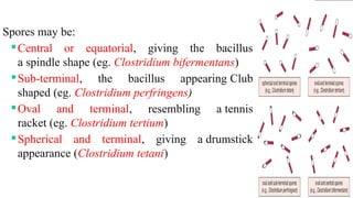
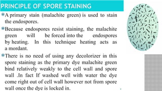
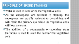
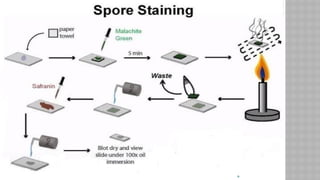
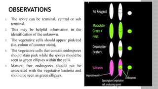
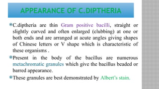
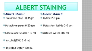
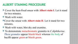
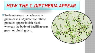
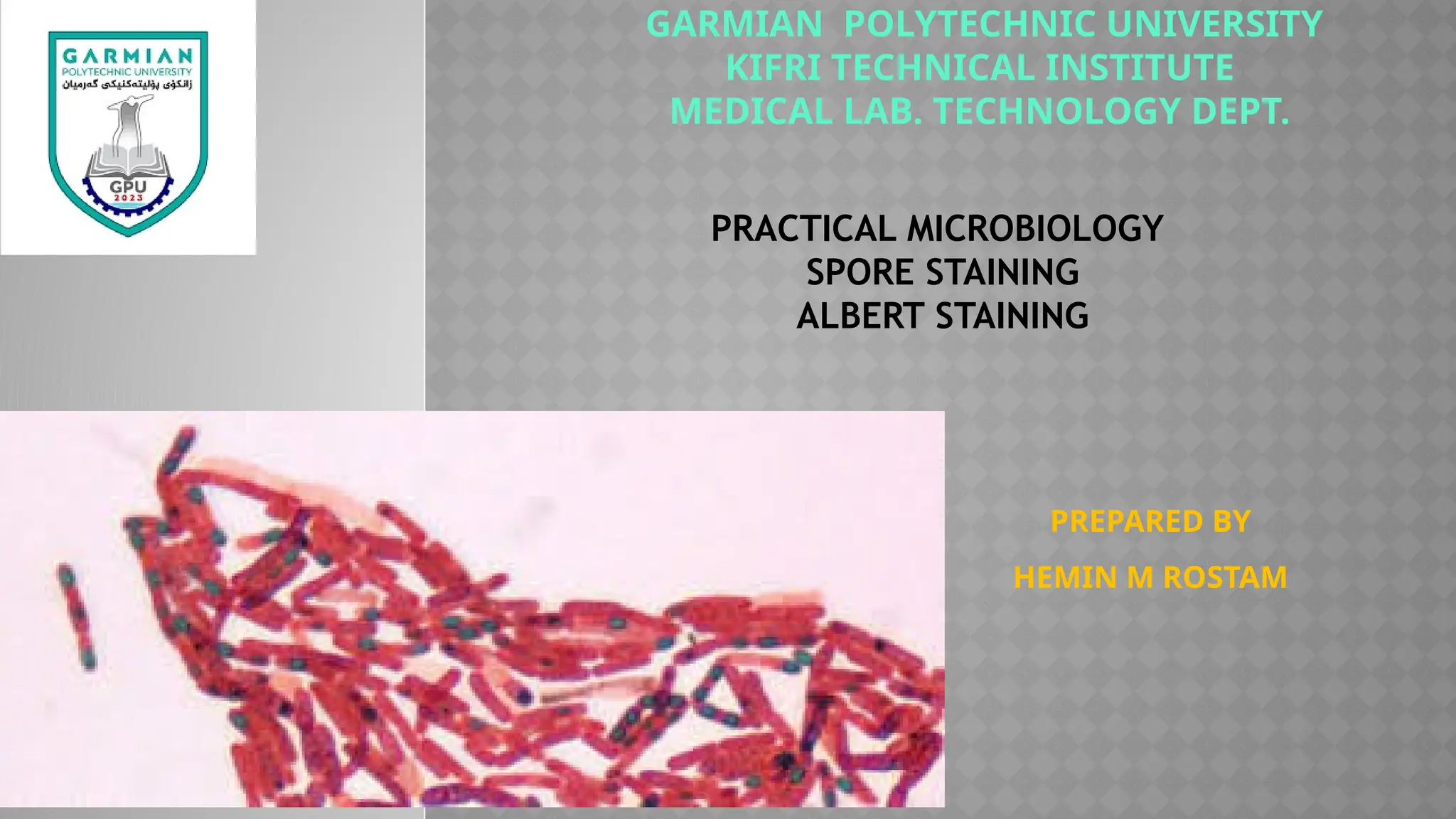
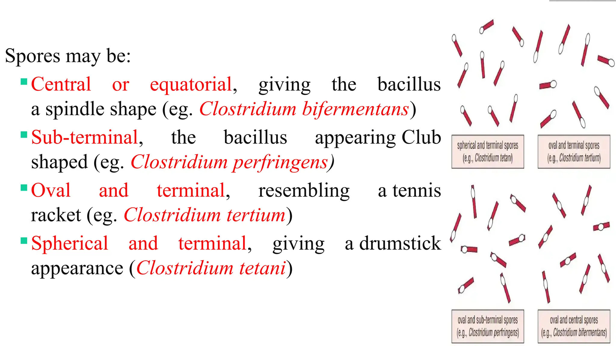
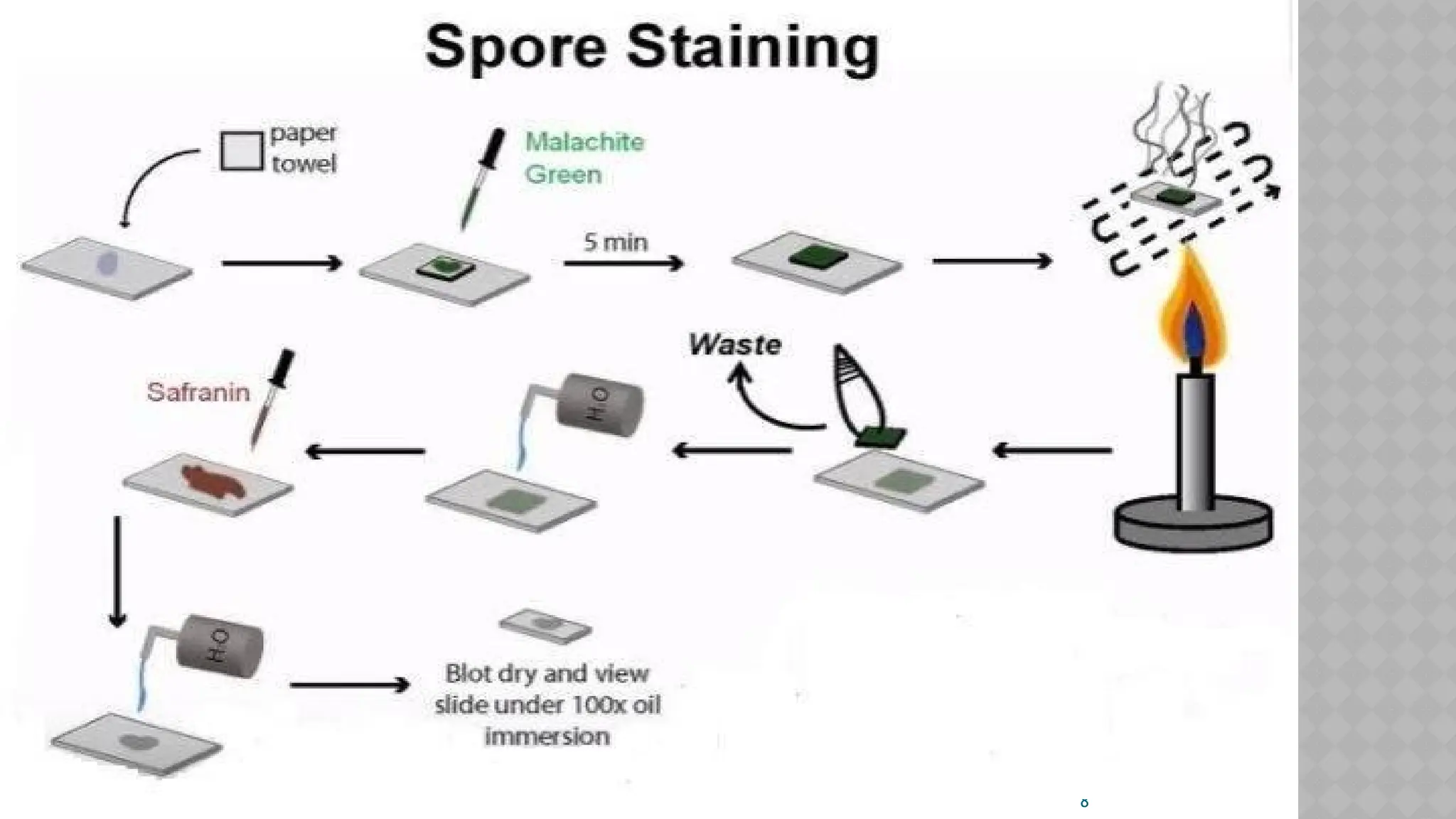
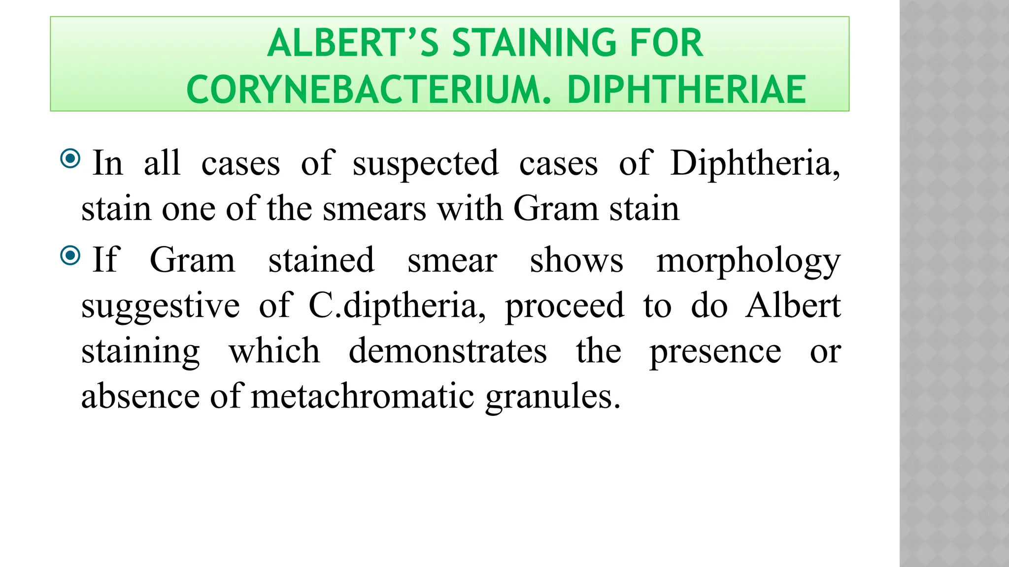
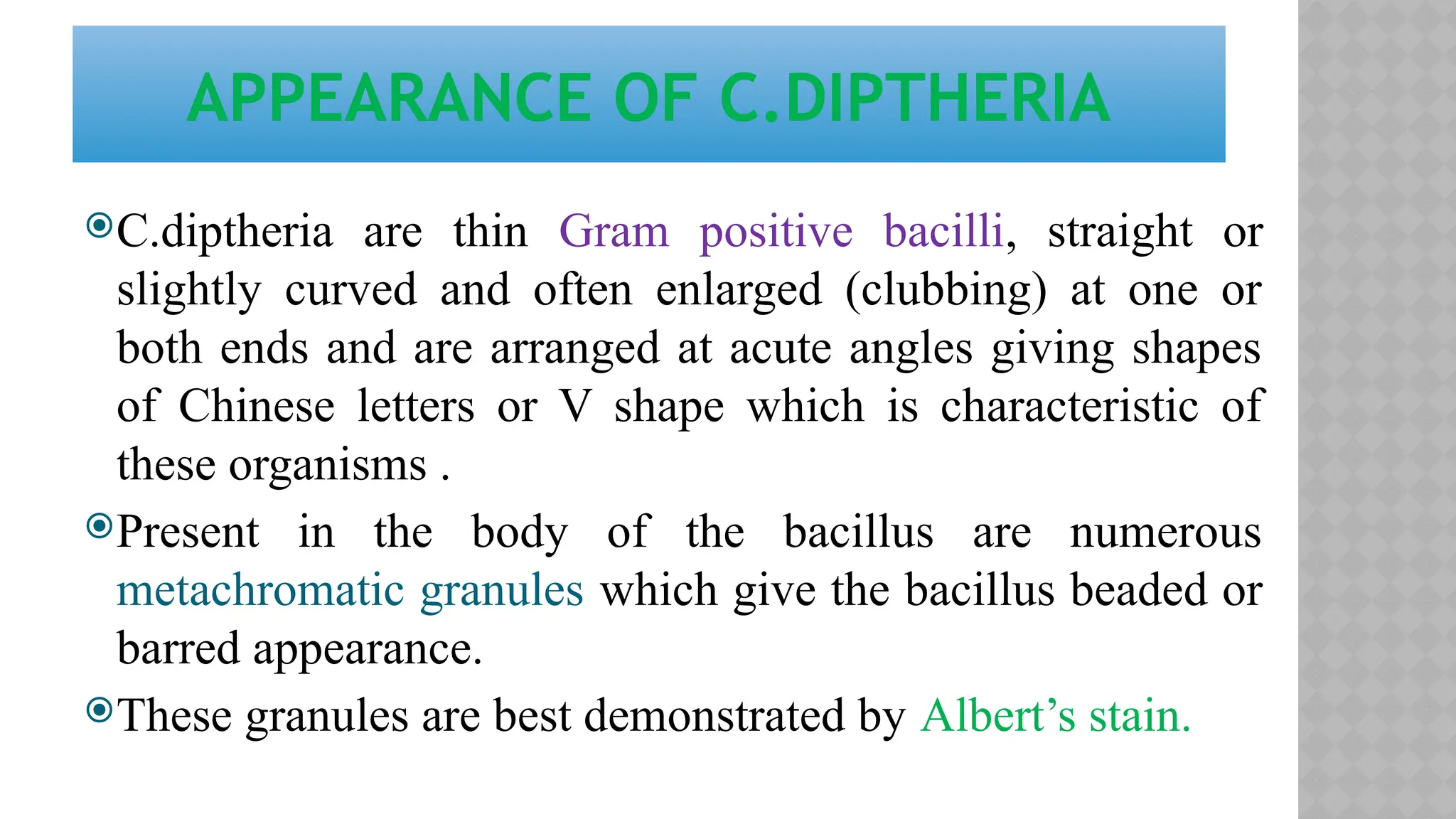
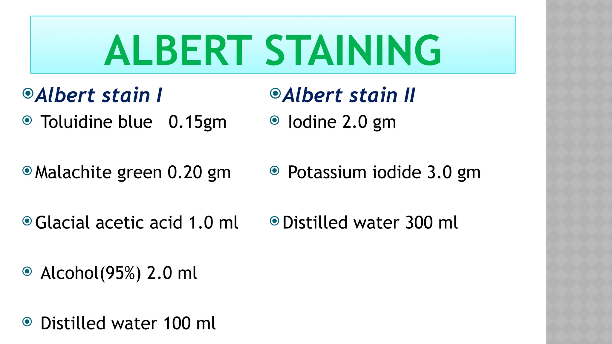
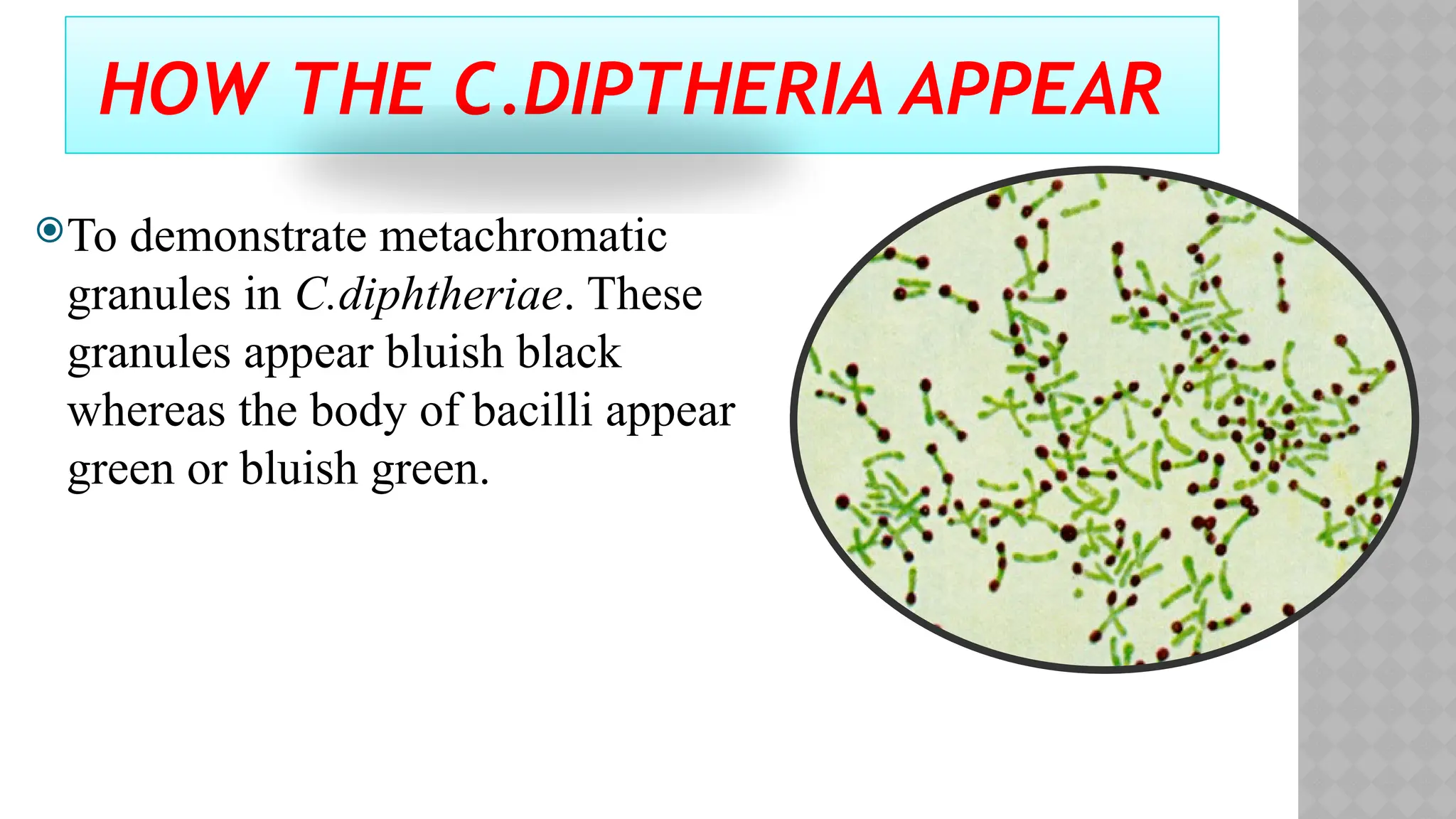
KEMBAR78
practical micro Lab5 presentation .pptx
PRACTICAL MICROBIOLOGY
SPORE STAINING
ALBERT STAINING
PREPARED BY
HEMIN M ROSTAM
GARMIAN POLYTECHNIC UNIVERSITY
KIFRI TECHNICAL INSTITUTE
MEDICAL LAB. TECHNOLOGY DEPT.
2
WHAT IS SPORE?
 Highly resistant, dormant structures (i.e. no metabolic activity)
formed in response to adverse environmental conditions.
 Help in the survival of the organisms during adverse environmental
conditions; do not have a role in reproduction.
 Spore formation (sporulation) occurs when nutrients, such as sources
of carbon and nitrogen are depleted.
 Are resistant to heat, dehydration, radiation and chemicals.
 Structurally and chemically more complex than the vegetative cell.
 The shape and the position of spores vary in different species and
can be useful for classification and identification purposes.
3
Spores may be:
Central or equatorial, giving the bacillus
a spindle shape (eg. Clostridium bifermentans)
Sub-terminal, the bacillus appearing Club
shaped (eg. Clostridium perfringens)
Oval and terminal, resembling a tennis
racket (eg. Clostridium tertium)
Spherical and terminal, giving a drumstick
appearance (Clostridium tetani)
4
PRINCIPLE OF SPORE STAINING
A primary stain (malachite green) is used to stain
the endospores.
Because endospores resist staining, the malachite
green will be forced into the endospores
by heating. In this technique heating acts as
a mordant.
There is no need of using any decolorizer in this
spore staining as the primary dye malachite green
bind relatively weakly to the cell wall and spore
wall .In fact If washed well with water the dye
come right out of cell wall however not from spore
wall once the dye is locked in.
5
PRINCIPLE OF SPORE STAINING
Water is used to decolorize the vegetative cells.
As the endospores are resistant to staining, the
endospores are equally resistant to de-staining and
will retain the primary dye while the vegetative cells
will lose the stain.
The addition of a counterstain or secondary stain
(safranin) is used to stain the decolorized vegetative
cells.
6
PROCEDURE
1. Slide,Loop,Spore-forming,bacteria,Malachitegreen,Safranin,Boiling
water bath/ Bunsen burner,Microscope,Immersion oil. Is required.
2. Prepare smears of organisms to be tested for presence of endospores
on a clean microscope slide and air dry it.
3. Heat fix the smear.
4. Place a small piece of blotting paper (absorbent paper) over the smear,
flood it with malachite green and place the slide (smear side up) on
a wire gauze on a ring stand.
5. Heat the slide gently till it starts to evaporate (either by putting the
slide on a staining rack that has been placed over a boiling water bath
or via Bunsen burner).
7
6. Remove the heat and reheat the slide as needed to keep the slide steaming for
about 3-5 minutes. as the paper begins to dry add a drop or two of malachite
green to keep it moist, but don’t add so much at one time that the temperature is
appreciably reduced.
7. Remove the blotting paper and allow the slide to cool to room temperature for 2
minutes.
8. Rinse the slide thoroughly with tap water.
9. Stain the smear with safranin for 2 minutes.
10. Rinse both side of the slide to remove the secondary stain and blot the slide/ air
dry.
11. Observe the bacteria under 1000x (oil immersion) total magnification.
8
9
OBSERVATIONS
1. The spore can be terminal, central or sub
terminal.
2. This may be helpful information in the
identification of the unknown.
3. The vegetative cells should appear pink/red
(i.e. colour of counter stain),
4. The vegetative cells that contain endospores
should stain pink while the spores should be
seen as green ellipses within the cells.
5. Mature, free endospores should not be
associated with the vegetative bacteria and
should be seen as green ellipses.
ALBERT’S STAINING FOR
CORYNEBACTERIUM. DIPHTHERIAE
 In all cases of suspected cases of Diphtheria,
stain one of the smears with Gram stain
 If Gram stained smear shows morphology
suggestive of C.diptheria, proceed to do Albert
staining which demonstrates the presence or
absence of metachromatic granules.
C.diptheria are thin Gram positive bacilli, straight or
slightly curved and often enlarged (clubbing) at one or
both ends and are arranged at acute angles giving shapes
of Chinese letters or V shape which is characteristic of
these organisms .
Present in the body of the bacillus are numerous
metachromatic granules which give the bacillus beaded or
barred appearance.
These granules are best demonstrated by Albert’s stain.
APPEARANCE OF C.DIPTHERIA
Albert stain I
 Toluidine blue 0.15gm
 Malachite green 0.20 gm
 Glacial acetic acid 1.0 ml
 Alcohol(95%) 2.0 ml
 Distilled water 100 ml
Albert stain II
 Iodine 2.0 gm
 Potassium iodide 3.0 gm
 Distilled water 300 ml
ALBERT STAINING
ALBERT STAINING PROCEDURE
 Cover the heat-fixed smear with Albert stain I. Let it stand
for two minutes.
 Wash with water.
Cover the smear with Albert stain II. Let it stand for two
minutes.
 Wash with water, blot dry and examine.
 To demonstrate metachromatic granules in C.diphtheriae.
These granules appear bluish black whereas the body of
bacilli appear green or bluish green.
HOW THE C.DIPTHERIA APPEAR
To demonstrate metachromatic
granules in C.diphtheriae. These
granules appear bluish black
whereas the body of bacilli appear
green or bluish green.
THANK YOU

practical micro Lab5 presentation .pptx

  • 1.
    PRACTICAL MICROBIOLOGY SPORE STAINING ALBERTSTAINING PREPARED BY HEMIN M ROSTAM GARMIAN POLYTECHNIC UNIVERSITY KIFRI TECHNICAL INSTITUTE MEDICAL LAB. TECHNOLOGY DEPT.
  • 2.
    2 WHAT IS SPORE? Highly resistant, dormant structures (i.e. no metabolic activity) formed in response to adverse environmental conditions.  Help in the survival of the organisms during adverse environmental conditions; do not have a role in reproduction.  Spore formation (sporulation) occurs when nutrients, such as sources of carbon and nitrogen are depleted.  Are resistant to heat, dehydration, radiation and chemicals.  Structurally and chemically more complex than the vegetative cell.  The shape and the position of spores vary in different species and can be useful for classification and identification purposes.
  • 3.
    3 Spores may be: Centralor equatorial, giving the bacillus a spindle shape (eg. Clostridium bifermentans) Sub-terminal, the bacillus appearing Club shaped (eg. Clostridium perfringens) Oval and terminal, resembling a tennis racket (eg. Clostridium tertium) Spherical and terminal, giving a drumstick appearance (Clostridium tetani)
  • 4.
    4 PRINCIPLE OF SPORESTAINING A primary stain (malachite green) is used to stain the endospores. Because endospores resist staining, the malachite green will be forced into the endospores by heating. In this technique heating acts as a mordant. There is no need of using any decolorizer in this spore staining as the primary dye malachite green bind relatively weakly to the cell wall and spore wall .In fact If washed well with water the dye come right out of cell wall however not from spore wall once the dye is locked in.
  • 5.
    5 PRINCIPLE OF SPORESTAINING Water is used to decolorize the vegetative cells. As the endospores are resistant to staining, the endospores are equally resistant to de-staining and will retain the primary dye while the vegetative cells will lose the stain. The addition of a counterstain or secondary stain (safranin) is used to stain the decolorized vegetative cells.
  • 6.
    6 PROCEDURE 1. Slide,Loop,Spore-forming,bacteria,Malachitegreen,Safranin,Boiling water bath/Bunsen burner,Microscope,Immersion oil. Is required. 2. Prepare smears of organisms to be tested for presence of endospores on a clean microscope slide and air dry it. 3. Heat fix the smear. 4. Place a small piece of blotting paper (absorbent paper) over the smear, flood it with malachite green and place the slide (smear side up) on a wire gauze on a ring stand. 5. Heat the slide gently till it starts to evaporate (either by putting the slide on a staining rack that has been placed over a boiling water bath or via Bunsen burner).
  • 7.
    7 6. Remove theheat and reheat the slide as needed to keep the slide steaming for about 3-5 minutes. as the paper begins to dry add a drop or two of malachite green to keep it moist, but don’t add so much at one time that the temperature is appreciably reduced. 7. Remove the blotting paper and allow the slide to cool to room temperature for 2 minutes. 8. Rinse the slide thoroughly with tap water. 9. Stain the smear with safranin for 2 minutes. 10. Rinse both side of the slide to remove the secondary stain and blot the slide/ air dry. 11. Observe the bacteria under 1000x (oil immersion) total magnification.
  • 8.
  • 9.
    9 OBSERVATIONS 1. The sporecan be terminal, central or sub terminal. 2. This may be helpful information in the identification of the unknown. 3. The vegetative cells should appear pink/red (i.e. colour of counter stain), 4. The vegetative cells that contain endospores should stain pink while the spores should be seen as green ellipses within the cells. 5. Mature, free endospores should not be associated with the vegetative bacteria and should be seen as green ellipses.
  • 10.
    ALBERT’S STAINING FOR CORYNEBACTERIUM.DIPHTHERIAE  In all cases of suspected cases of Diphtheria, stain one of the smears with Gram stain  If Gram stained smear shows morphology suggestive of C.diptheria, proceed to do Albert staining which demonstrates the presence or absence of metachromatic granules.
  • 11.
    C.diptheria are thinGram positive bacilli, straight or slightly curved and often enlarged (clubbing) at one or both ends and are arranged at acute angles giving shapes of Chinese letters or V shape which is characteristic of these organisms . Present in the body of the bacillus are numerous metachromatic granules which give the bacillus beaded or barred appearance. These granules are best demonstrated by Albert’s stain. APPEARANCE OF C.DIPTHERIA
  • 12.
    Albert stain I Toluidine blue 0.15gm  Malachite green 0.20 gm  Glacial acetic acid 1.0 ml  Alcohol(95%) 2.0 ml  Distilled water 100 ml Albert stain II  Iodine 2.0 gm  Potassium iodide 3.0 gm  Distilled water 300 ml ALBERT STAINING
  • 13.
    ALBERT STAINING PROCEDURE Cover the heat-fixed smear with Albert stain I. Let it stand for two minutes.  Wash with water. Cover the smear with Albert stain II. Let it stand for two minutes.  Wash with water, blot dry and examine.  To demonstrate metachromatic granules in C.diphtheriae. These granules appear bluish black whereas the body of bacilli appear green or bluish green.
  • 14.
    HOW THE C.DIPTHERIAAPPEAR To demonstrate metachromatic granules in C.diphtheriae. These granules appear bluish black whereas the body of bacilli appear green or bluish green.
  • 15.