Downloaded 271 times


This document describes the process of spore staining to differentiate bacterial spores from vegetative cells. It explains that spores are dormant, resistant structures formed by bacteria during adverse environmental conditions for survival. The spore staining technique uses malachite green as the primary stain for spores and safranin as the counterstain for vegetative cells. Heat is applied to help the malachite green penetrate the spore walls. Vegetative cells are decolorized but spores retain the green stain. This allows spores and vegetative cells to be distinguished microscopically.
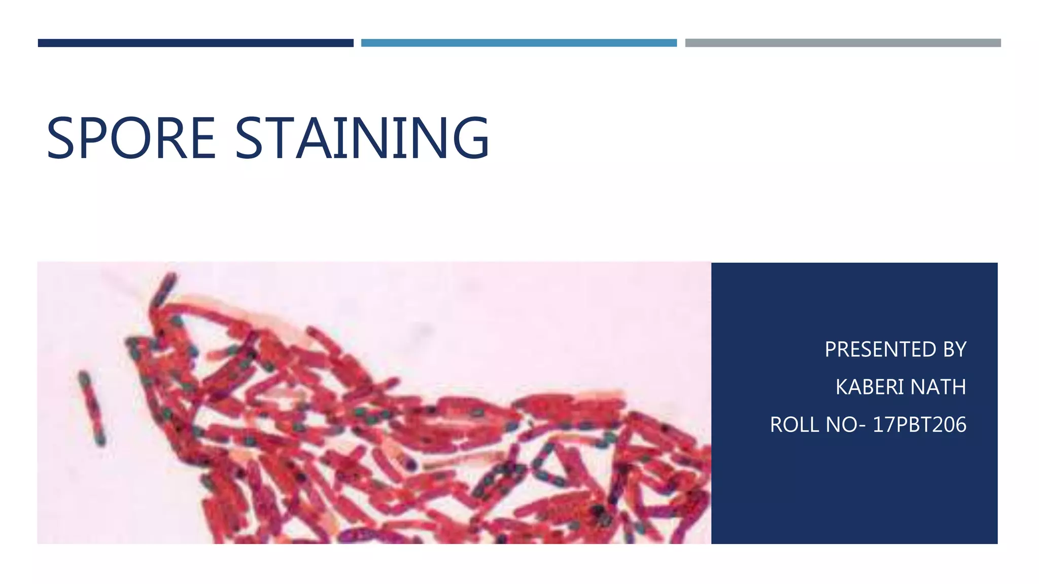
Introduction and presentation details by Kaberi Nath on spore staining.
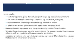
Definition of spores, types (central, sub-terminal, terminal), and germination process.
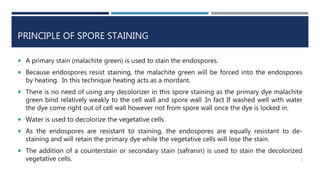
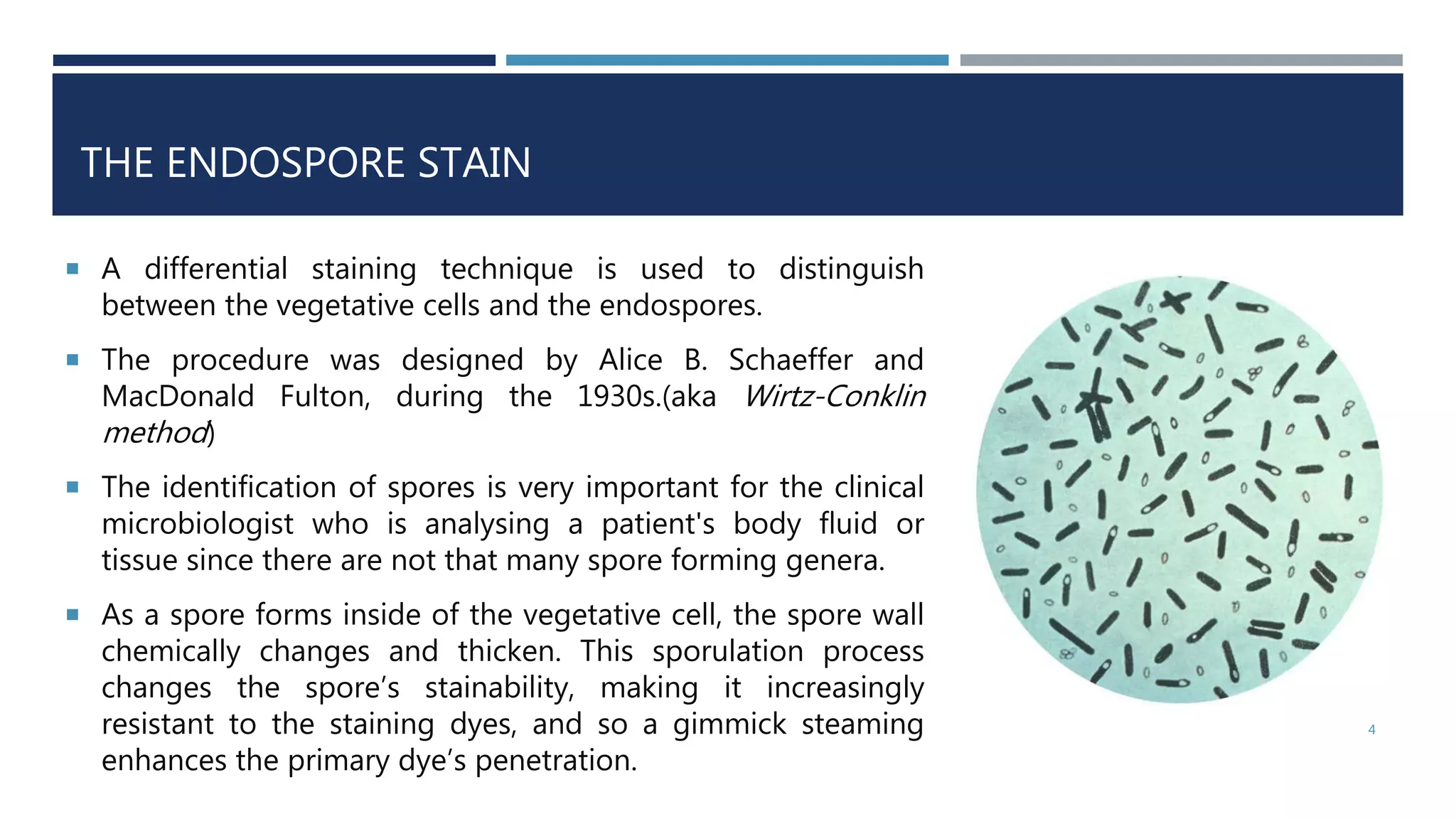
Overview of the differential staining technique, its importance, and principle using malachite green.
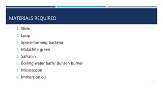
List of materials required for spore staining including slide, bacteria, and staining agents.
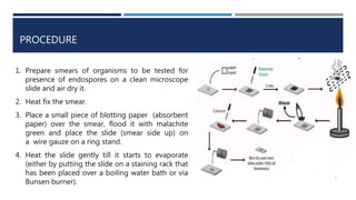
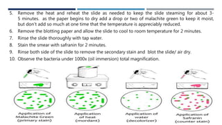
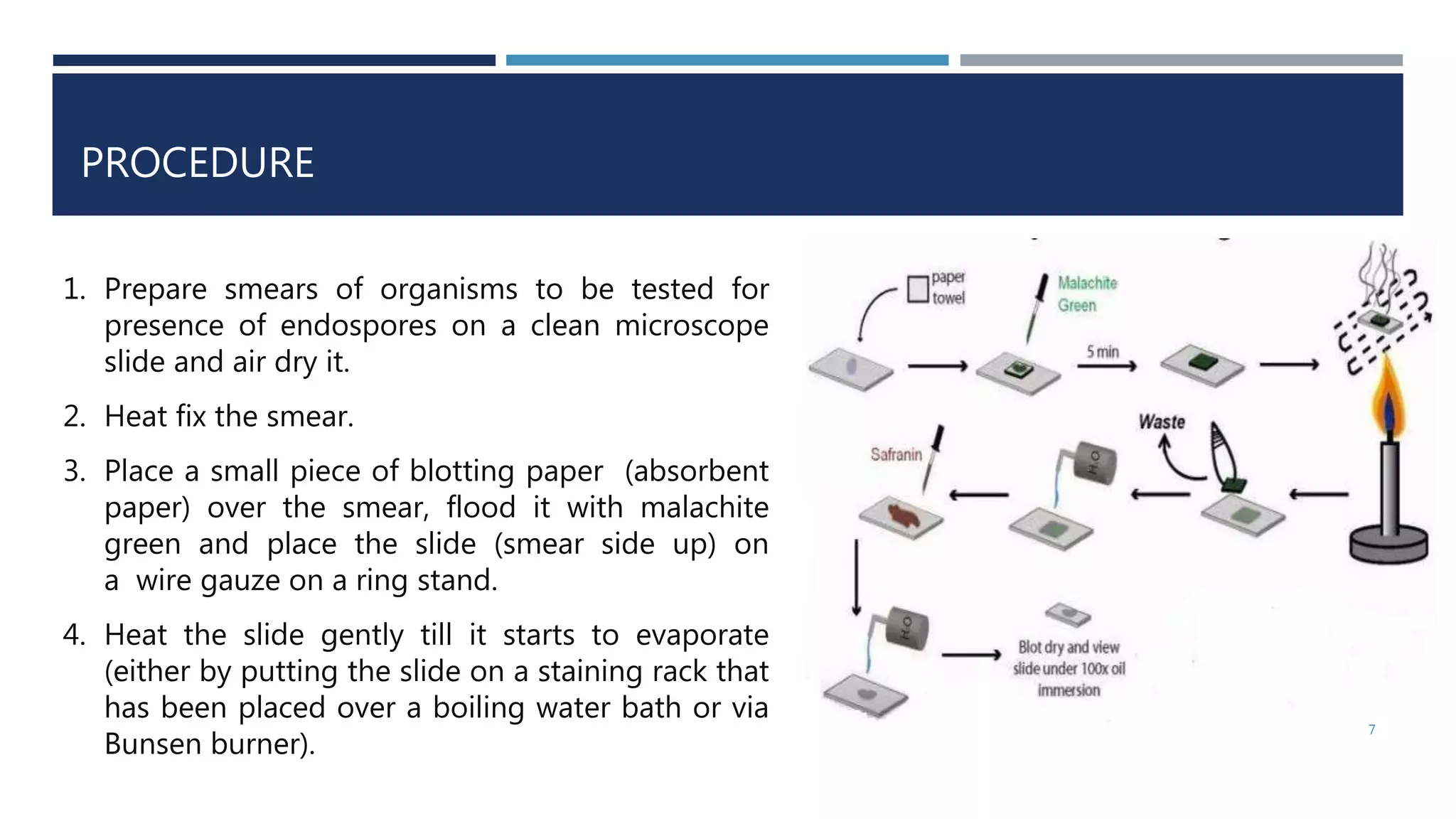
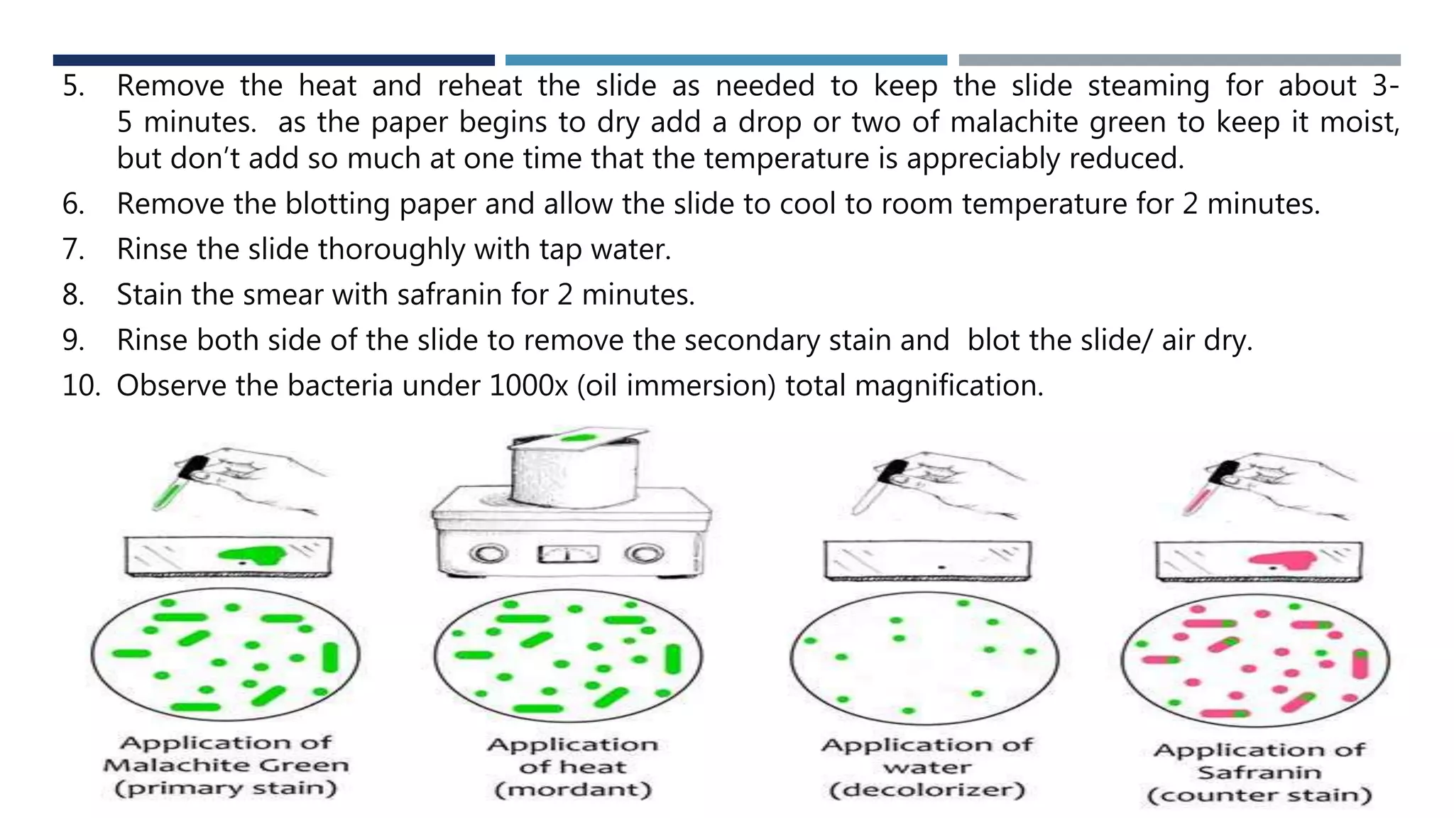
Step-by-step procedure for spore staining, including heat fixing and staining techniques.
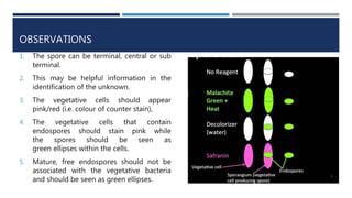
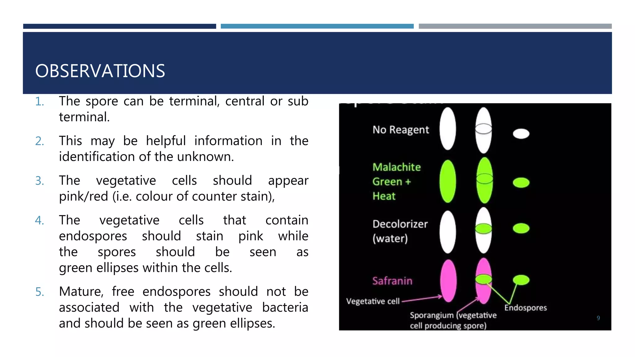
Expected results and observations post-staining; identification of spores and vegetative cells.
Citations and resources for the study and method of spore staining.
Blank slide without any content.


